Clark's Pocket Handbook for Radiographers
Hodder Arnold (Verlag)
978-0-340-93993-2 (ISBN)
- Titel ist leider vergriffen;
keine Neuauflage - Artikel merken
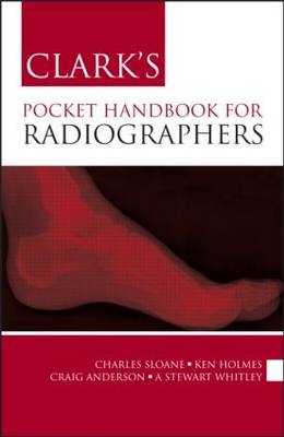
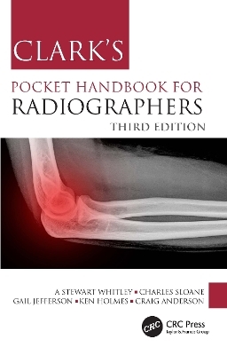
This pocket-sized guide, drawn from the twelfth edition of Clark's Positioning in Radiography, provides clear and practical advice to help radiographers in their day-to-day work. The authors considered that it is important for radiographers and students to have access to an additional text available in a "pocket" format which is easily transportable and convenient to use during everyday radiographic practice.
Designed for rapid reference, it covers how to position the patient and the central ray, describes the essential image characteristics, and illustrates each radiographic projection with a positioning photograph and a radiograph.
The authors have included a range of additional information new to this text. This includes a protocol for evaluating images (the "10-point plan") and a range of general advice for undertaking procedures in a professional and efficient manner. The book also includes basic information in relation to some non-imaging diagnostic tests, common medical terminology, and abbreviations. This is designed to help readers gain a better understanding of the diagnostic requirements and role of particular imaging procedures from the information presented in X-ray requests. In addition, the book discusses image evaluation, medical abbreviations, relevant normal blood values, and radiation protection.
Together with key points, this information helps the radiographer achieve the ideal image result.
Charles Sloane MSc DCR DRI, Principal Lecturer and Radiography Course Leader, University of Cumbria, Lancaster, UK Ken Holmes MSc TDCR DRI Cert CI, Senior Lecturer, School of Medical Imaging Sciences, University of Cumbria, Lancaster, UK Craig Anderson MSc BSc, Clinical Tutor, X-ray Department, Furness General Hospital, Cumbria, UK A Stewart Whitley FCR TDCR HDCR FETC, Radiology Advisor, UK Radiology Advisory Services Ltd, Preston, UK
Key Aspects of Radiographic Practice
Anatomical Terminology
Positioning Terminology
Projection Terminology
General Considerations for the Conduct of Radiographic Examinations
Patient identification and Consent
Justification of Examination
Radiation protection
Pregnancy
Evaluating Images: The 10-Point Plan
Examination Timeline
Guidelines for the Assessment of Trauma
Theatre Radiography
Radiographic Projections
Acromioclavicular joint
Abdomen
Ankle: Lateral
Calcanium
Cervical Spine
Chest
Clavicle
Coccyx Lat
Elbow
Facial Bones
Femur
Fingers
Foot
Forearm
Hand
Hip
Humerus
Knee
Lumbar Spine
Mandible
Orbits
OPG
Pelvis
Sacroiliac Joints
Sacrum
Scaphoid
Shoulder
Sinuses
Skull
Sternum
Thumb
Thoracic Spine
Thumb: Lateral
Tibia & Fibula
Toes
Wrist
Zygomatic arches
Useful Information for Radiographic Practice
Non Imaging Diagnostic Tests
Medical Terminology
Abbreviations
| Erscheint lt. Verlag | 30.4.2010 |
|---|---|
| Zusatzinfo | 22 b/w line drawings; 200 b/w halftones |
| Verlagsort | London |
| Sprache | englisch |
| Maße | 129 x 198 mm |
| Gewicht | 340 g |
| Themenwelt | Medizin / Pharmazie ► Gesundheitsfachberufe ► MTA - Radiologie |
| Medizin / Pharmazie ► Medizinische Fachgebiete ► Radiologie / Bildgebende Verfahren | |
| ISBN-10 | 0-340-93993-1 / 0340939931 |
| ISBN-13 | 978-0-340-93993-2 / 9780340939932 |
| Zustand | Neuware |
| Haben Sie eine Frage zum Produkt? |
aus dem Bereich


