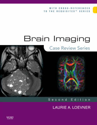
Brain Imaging: Case Review Series
Mosby (Verlag)
978-0-323-03179-0 (ISBN)
- Titel erscheint in neuer Auflage
- Artikel merken
This volume in the best-selling "Case Review" series uses hundreds of case studies to challenge your knowledge of a full range of topics in brain imaging. With 170 brand new cases, new coverage of MRA, CTA, MR spectroscopy and multi-detectors and over 600 brilliant images, this is your ideal concise, economical, and user-friendly tool for self assessment in this specialty!
Utilizes case studies organized into "Opening Round," "Fair Game," and "Challenge" sections, so you can test yourself at varying difficulty levels.
Provides at-a-glance review/self-testing of brain imaging cases ideal for preparing for the boards in brain imaging, the CAQ exam for neuroradiology or for the general radiologist ready for re-certification.
Mimics the official exam formats and daily practice environment by giving you cases/images as unknowns with three to four questions; then, on the flip side of the page, diagnosis, answers to the questions, additional commentary, and references to the corresponding volume in Elsevier's popular Requisites Series.
Includes 600 state of the art images to effectively compliment and support the text and provide a clear picture of what you can expect, both in test-taking and in practice.
Uses randomly organized cases so you can test yourself without the aid of logical organization by anatomy or disease type.
Includes 170 new cases and over 50 new diagnoses so you can keep pace with the latest developments.
Includes a greater emphasis on differential diagnosis.
Adds coverage of MRA, CTA, MR spectroscopy and multi-detectors to keep you completely current.
Provides all new images for existing entities.
Adds cutting-edge coverage of neuro-imaging including spectroscopy, CTA, MRA, Functional imaging, tractography, perfusion and diffusion.
1 Acute Anterior Cerebral Artery Infarct
2 Cerebral Arteriovenous Malformation
3 Hemorrhagic Metastases - Melanoma
4 Penetrating Brain Injury - Bow and Arrow Injury
5 Orbital Pseudotumor
6 Acute Epidural Hematoma and Associated Linear Fracture of the Frontal Bone
7 Pyogenic Brain Abscess
8 Chiari II Malformation - with Associated Anomalies
9 Acute Hydrocephalus Secondary to Meningitis
10 Pineal Cyst
11 Hemorrhagic Contusions - Closed Head Injury
12 Juvenile Pilocytic Astrocytoma of the Cerebellum
13 Fibrous Dysplasia
14 Acute Middle Cerebral Artery Stroke: "Hyperdense MCA and Insular Ribbon Sign
15 Multiple Sclerosis
16 Meningioma
17 Acute Actively bleeding Subdural hematoma - Subfalcine Herniation and Stroke
18 Acute Subarachnoid Hemorrhage - Rupture of an Anterior Communicating Artery Aneurysm
19 Embolic Infarcts (Acute and Subacute)- Atrial Fibrillation
20 Vestibular Schwannoma of the Internal Auditory Canal
21 Orbital Cellulitis and Abscess
22 Acute Hypertensive Thalamic Hemorrhage
23 Infiltrating Astrocytoma - Low Grade
24 Arachnoid Cyst
25 Retinoblastoma
26 Subdural Empyema - Complicated by Cerebritis
27 Glioblastoma Multiforme of the Corpus Callosum--"Butterfly Glioma"
28 Virchow-Robin Perivascular Space
29 Agenesis of the Corpus Callosum
30 Nonaccidental Trauma--Child Abuse
31 Cerebellar Hemangioblastoma
32 Calvarial Metastases--Breast Carcinoma
33 Chronic Anemia - Diffuse Replacement of the Fat in the Calvarial Marrow
34 Giant Aneurysm - Middle Cerebral Artery
35 Bilateral Subacute Subdural Hematomas
36 Parafalcine Meningioma Invading the Superior Sagittal Sinus
37 Hemorrhagic Venous Infarction
38 Vertebral Artery Dissection - Spontaneous
39 Spontaneous Cerebral Hematoma - Ruptured Cerebral Arteriovenous Malformation
40 Vascular Infundibulum - Posterior Communicating Artery
41 Closed Head Injury - Diffuse Axonal Injury
42 Glioblastoma Multiforme - Subependymal Spread
43 Amyloid Angiopathy
44 Mesial Temporal Sclerosis
45 Intraventricular Meningioma
46 Suprasellar Germinoma
47 Mineral Deposition in the Basal Ganglia on T1W Imaging - Abnormal Calcium/Phosphate Metabolism
48 Herpes Simplex Encephalitis - Type 1
49 Primary Orbital Lymphoma
50 Basilar Meningitis and Encephalitis - Tuberculosis
FAIR GAME
51 Dural Metastases - Breast Carcinoma
52 Aqueductal Stenosis
53 Rathke's Cleft Cyst
54 Optic Neuritis (Demyelinating Disease)
55 Ocular trauma - Lens dislocation; Globe Rupture
56 Developmental Venous Anomaly
57 Fibrous Dysplasia of the Calvaria
58 Cavernous Sinus Mass-Meningioma
59 Pyogenic Ventriculitis With Acute Hydrocephalus
60 Lesions of the Pituitary Stalk and Hypothalamus: Case A Sarcoidosis, Case B Langerhans Histiocytosis (EG)
61 Nodular Subependymal Heterotopia
62 Basilar Meningitis - Sarcoidosis
63 Pituitary Microadenoma
64 Multiple Sclerosis - Marburg Type
65 Transtentorial Herniation
66 Epidermoid Cyst - Cerebellopontine Cistern
67 Cavernous Malformation (Cavernous Hemangioma, Occult Cerebrovascular Malformation)
68 Persistent Trigeminal Artery
69 Colloid Cyst
70 Cavum Septum Pellucidum and Cavum Vergae
71 Toxoplasmosis Infection in Acquired Immunodeficiency Syndrome
72 Encephalocele - Developmental
73 Cysticercosis of the Central Nervous System
74 Facial nerve - Inflammation (Viral)
75 MR Tractography - Preoperative Mapping of the Corticospinal Tracts for GBM Resection
76 Right Posterior Inferior Cerebellar Artery Aneurysm Rupture
77 Ganglioglioma
78 Paget's Disease - Osteitis Deformans
79 Fibromuscular Dysplasia - Complicated by Acute Stroke
80 Carbon Monoxide Poisoning
81 Lateral Medullary Syndrome - Wallenberg's
82 Wallerian Degeneration
83 Dandy-Walker Malformation
84 Thyroid Ophthalmopathy - with Optic Nerve Compression in Orbital Apex
85 Bilateral Frontal Sinus Mucoceles
86 Suprasellar Cistern Arachnoid Cyst
87 Fourth Ventricular Neoplasms - Choroid Plexus Papilloma, and Ependymoma
88 Multiple Cavernous Malformations - Familial Pattern
89 Vein of Galen Aneurysm
90 Tuberous Sclerosis (Bourneville's Disease)
91 Craniosynostosis-Metopic Suture
92 Glioma of the Tectum (Quadrigeminal Plate)
93 Craniopharyngioma - Recurrent Adamantinomatous Type
94 Intradiploic Epidermoid Cyst
95 Porencephalic Cyst
96 Subependymal Heterotopia
97 Acute Middle Cerebral Artery Stroke - MR
98 Hemangioblastoma
99 Posterior Circulation Ischemia due to Subclavian Steal
100 CNS Vasculitis - Cerebral Hemorrhage and Infarcts
101 Optic Nerve Meningioma
102 Peri-pineal Meningioma
103 Air Embolism with Acute Right Cerebral Infarct
104 Venous Sinus Thrombosis Complicating Otomastoiditis
105 Intraventricular Hemorrage
106 Pituitary Macroadenoma - in Left Cavernous Sinus
107 Lymphoma of Occipital Condyle and C1
108 Central Nervous System Sarcoidosis
109 Normal-Pressure Hydrocephalus
110 Lipomas Associated with the Corpus Callosum
111 Sinonasal Undifferentiated Carcinoma with Intracranial Extension
112 Intracerebral Rupture of a Posterior Communicating Artery Aneurysm
113 Gadolinium-Associated Nephrogenic Systemic Fibrosis (Part I)
114 Gadolinium-Associated Nephrogenic Systemic Fibrosis (Part II)
115 Fenestration of the Basilar Artery
116 Moyamoya Disease
117 Focal Cortical Dysplasia
118 Acute Sinusitis
119 Anterior Clinoiditis and Mucocele Formation Complicating Sinusitis
120 Spontaneous Intracranial Hypotension
121 Multiple Meningiomas Following Remote Whole Brain Irradiation
122 Fahr Disease (Familial Cerebrovascular Ferrocalcinosis)
123 Central Nervous System Lymphoma--Immunocompetent Patient
124 Alzheimer's Disease
125 Sturge-Weber Syndrome (Encephalotrigeminal Angiomatosis)
126 Ectopic Posterior Pituitary and Nonvisualization of the Infundibulum - Hypopituitarism
127 Acute Toxic Demyelination - Carbon Monoxide Intoxication
128 Neurofibromatosis Type 1 - Pilocytic Astrocytoma of Optic Pathway
129 Lymphoma Involving the Circumventricular Organs
130 Nonaneurysmal Perimesencephalic Subarachnoid Hemorrhage
131 Radiation Necrosis Following Treatment of High-Grade Glioma
132 Radiation Necrosis After XRT for Nasopharyngeal Cancer
133 Internal Carotid Artery Dissection
134 Tension Pneumocephalus
135 Cryptococcosis
136 Subarachnoid Seeding--Leptomeningeal Carcinomatosis
137 Brainstem Astrocytoma
138 Thrombosis of a Developmental Venous Malformation - Postpartum
139 Textiloma or Gossypiboma - Retained Surgical Sponge
140 Petrous Apex Cholesterol Granuloma
141 Hamartoma of the Hypothalamus
142 Hodgkin Disease - Dural Based
143 Clival Chordoma
144 Perineural Spread of Skin Cancer - Basal Cell Carcinoma
145 Central Nervous System Lyme Disease
146 Reversible Posterior Leukoencephalopathy (RPL) - PRES
147 Fibrous Dysplasia of the Temporal Bone
148 Neuroblastoma - Metastases to the Cranium
149 Cystic Meningioma - with Cellular Atypia
150 Global Anoxic Brain Injury
CHALLENGER
151 Lobar Holoprosencephaly
152 Olivopontocerebellar Degeneration
153 Band Heterotopia-Pachygyria
154 Huntington's Disease
155 Meningocele/Pseudomeningocele of the Skull Base (Petrous Apex)
156 Right Thalamic Glioblastoma Multiforme
157 Multiple Sclerosis and Glioblastoma Multiforme
158 Adrenoleukodystrophy
159 Medulloblastoma
160 Subacute Middle Cerebral Artery Territory Stroke Mimicking a Bone Metastasis
161 Cytomegalovirus Meningitis and Ependymitis in a Patient with AIDs
162 Upper Brainstem and Thalamic Cavernous Malformation
163 Capillary Telangiectasia and Developmental Venous Anomaly of the Brain Stem
164 Central Neurocytoma
165 Marchiafava-Bignami Syndrome
166 Metastatic Carcinoid to the Orbital Extraocular Muscles
167 Joubert's Syndrome
168 Systemic Non-hodgkin Lymphoma
169 Abnormal Iron Deposition in Deep Gray Matter Nuclei
170 Herpes Encephalopathy - HHV-6 Following Organ Transplantation
171 Jacob-Creutzfeldt Disease
172 Orbital Lymphangioma
173 Carotid-Cavernous Fistula - Part I
174 Carotid-Cavernous Fistula - Part II
175 Gliomatosis Cerebri
176 Canavan's Disease
177 Neurofibromatosis Type 2
178 Metastasis to the Cerebellopontine Angle - Ocular Melanoma
179 Retained Gadolinium - Renal Failure
180 Amyotrophic Lateral Sclerosis - Lou Gehrig's Disease
181 High Grade Anaplastic Astrocytoma
182 Wilson's Disease
183 Pleomorphic Xanthoastrocytoma
184 Primary Leptomeningeal Melanosis
185 Dural Arteriovenous Fistula (Part I)
186 Dural Arteriovenous Fistula (Part II)
187 Fetal Schizencephaly
188 Wernicke Encephalopathy
189 Ruptured Mycotic Aneurysm
190 Osmotic Demyelination
191 Rosai-Dorfman Disease - Benign Sinus Histiocytosis
192 Cystic Necrotic Brain Mass - Part I
193 Cystic Necrotic Brain Mass Part II- Metastatic Adenocarcinoma of the Lung
194 Rhombencephalosynapsis
195 Progressive Multifocal Leukoencephalopathy
196 Sphenoid Dysplasia and Brain Dysplasia- Neurofibromatosis Type 1 (von Recklinghausen's Disease)
197 Panhypopituitarism with Absence of the Infundibulum
198 Progressive External Ophthalmoplegia Syndrome - Due to Mitochondrial Defect
199 Limbic Encephalitis - Paraneoplastic Syndrome (Small Cell Lung Carcinoma)
200 von Hippel-Lindau Disease
| Erscheint lt. Verlag | 22.3.2021 |
|---|---|
| Reihe/Serie | Case Review Series |
| Zusatzinfo | Approx. 640 illustrations; Illustrations |
| Verlagsort | St Louis |
| Sprache | englisch |
| Maße | 216 x 276 mm |
| Gewicht | 1130 g |
| Themenwelt | Medizin / Pharmazie ► Medizinische Fachgebiete ► Neurologie |
| Medizinische Fachgebiete ► Radiologie / Bildgebende Verfahren ► Radiologie | |
| Schlagworte | Bildgebende Verfahren (Medizin) • Gehirn |
| ISBN-10 | 0-323-03179-X / 032303179X |
| ISBN-13 | 978-0-323-03179-0 / 9780323031790 |
| Zustand | Neuware |
| Haben Sie eine Frage zum Produkt? |
aus dem Bereich


