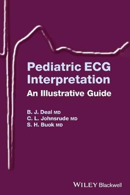
Pediatric ECG Interpretation
An Illustrative Guide
Seiten
2004
Wiley-Blackwell (Verlag)
978-1-4051-1730-2 (ISBN)
Wiley-Blackwell (Verlag)
978-1-4051-1730-2 (ISBN)
Illustrating many of the ECG patterns a pediatric practitioner is likely to encounter, this title presents those illustrations with interpretations in several categories: normal children of various ages, acquired abnormalities such as hypertrophy or electrolyte disorders, and common congenital heart disease lesions.
Pattern recognition is an important learning tool in the interpretation of ECGs. Unfortunately, until faced with a patient with an arrhythmia or structural heart disease, pediatric practitioners generally receive limited exposure to ECGs. The ability to clearly distinguish an abnormal ECG pattern from a normal variant in an emergency situation is an essential skill, but one that many pediatricians feel ill-prepared to utilize confidently. In Pediatric ECG Interpretation: An Illustrative Guide, Drs. Deal, Johnsrude and Buck aim to address this issue by illustrating many of the ECG patterns a pediatric practitioner is likely to encounter.
ECG illustrations with interpretations are presented in several categories: normal children of all ages, acquired abnormalities such as hypertrophy or electrolyte disorders, and common congenital heart disease lesions. Later sections cover bradycardia, supraventricular and ventricular arrhythmias, and a basic section on pacemaker ECGs. Simple techniques used to interpret mechanisms of arrhythmias are described as a resource for practitioners in cardiology, adult electrophysiology, or pediatrics who may not have a readily accessible resource for these ECG examples.
Material hosted at http://wiley.mpstechnologies.com/wiley/BOBContent/searchLPBobContent.do can be used:
1 as a self-evaluation tool for interpretation of ECGs
2 as a teaching reference for Cardiology fellows, residents, and house staff
3 as an invaluable resource for the Emergency Room physician or pediatrician who might obtain an ECG on a pediatric patient
Dr. Barbara J. Deal, MD. Associate Professor of Pediatrics, Northwestern University Medical School Dr. Christopher L. Johnsrude, MD. Associate Professor of Pediatrics, Director of Electrophysiology, University of Louisville School of Medicine
Introduction. Normal ECGs.
Abnormal ECGs.
Acquired Heart Disease.
Congenital Heart Disease.
Bradycardia and Conduction Defects.
Supraventricular Tachycardia.
Ventricular Arrhythmias.
Pacemakers.
Appendix 1: Age-related normal ECG values in children.
Appendix 2: Criteria for distinguishing VT from SVT.
Appendix 3: Location of accessory atrioventricular connection using initial delta wave polarity.
Appendix 4: Indications for pacing in childhood.
Index
| Erscheint lt. Verlag | 1.6.2004 |
|---|---|
| Verlagsort | Hoboken |
| Sprache | englisch |
| Maße | 152 x 229 mm |
| Gewicht | 386 g |
| Themenwelt | Medizinische Fachgebiete ► Innere Medizin ► Kardiologie / Angiologie |
| Medizin / Pharmazie ► Medizinische Fachgebiete ► Pädiatrie | |
| Medizin / Pharmazie ► Medizinische Fachgebiete ► Radiologie / Bildgebende Verfahren | |
| ISBN-10 | 1-4051-1730-3 / 1405117303 |
| ISBN-13 | 978-1-4051-1730-2 / 9781405117302 |
| Zustand | Neuware |
| Haben Sie eine Frage zum Produkt? |
Mehr entdecken
aus dem Bereich
aus dem Bereich
in Fällen, Fragen und Antworten
Buch | Softcover (2024)
Urban & Fischer in Elsevier (Verlag)
89,00 €
Diagnostik und interventionelle Therapie | 2 Bände
Buch (2024)
Deutscher Ärzteverlag
349,99 €


