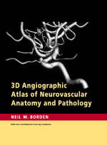
3D Angiographic Atlas of Neurovascular Anatomy and Pathology
Cambridge University Press (Verlag)
978-0-521-85684-3 (ISBN)
The 3D Angiographic Atlas of Neurovascular Anatomy and Pathology is the first atlas to present neurovascular information and images based on catheter 3D rotational angiographic studies. The images in this book are the culmination of work done by Neil M. Borden over several years using one of the first 3D neurovascular angiographic suites in the United States. With the aid of this revolutionary technology, Dr Borden has performed numerous diagnostic neurovascular angiographic studies as well as endovascular neurosurgical procedures. The spectacular 3D images he obtained are extensively labeled and juxtaposed with conventional 2D angiograms for orientation and comparison. Anatomical color drawings and concise descriptions of the major intracranial vascular territories further enhance understanding of the complex cerebral vasculature.
Neil M. Borden, MD, is a board-certified neuroradiologist who has been practising for 20 years. He completed a neuroradiology fellowship at the Neurological Institute of New York at Columbia Presbyterian Medical Center and a two-year fellowship in endovascular neurosurgery at the Barrow Neurological Institute in Phoenix, Arizona. Dr Borden is a senior member of the American Society of Neuroradiology and is currently practising at the Cleveland Clinic Foundation in Cleveland, Ohio.
Introduction; 1. Technique of 3D rotational angiography; 2. Color illustrations of vasculature; 3. The aortic arch; 4. Cervical vasculature; 5. Intracranial carotid circulation: anterior circulation; 6. Intracranial vertebral basilar circulation: posterior circulation; 7. Intracranial venous circulation; 8. The circle of Willis.
| Erscheint lt. Verlag | 4.12.2006 |
|---|---|
| Zusatzinfo | 593 Halftones, unspecified; 16 Line drawings, color |
| Verlagsort | Cambridge |
| Sprache | englisch |
| Maße | 220 x 287 mm |
| Gewicht | 1025 g |
| Themenwelt | Medizinische Fachgebiete ► Innere Medizin ► Kardiologie / Angiologie |
| Medizin / Pharmazie ► Medizinische Fachgebiete ► Neurologie | |
| Medizinische Fachgebiete ► Radiologie / Bildgebende Verfahren ► Radiologie | |
| Studium ► 1. Studienabschnitt (Vorklinik) ► Anatomie / Neuroanatomie | |
| ISBN-10 | 0-521-85684-1 / 0521856841 |
| ISBN-13 | 978-0-521-85684-3 / 9780521856843 |
| Zustand | Neuware |
| Informationen gemäß Produktsicherheitsverordnung (GPSR) | |
| Haben Sie eine Frage zum Produkt? |
aus dem Bereich


