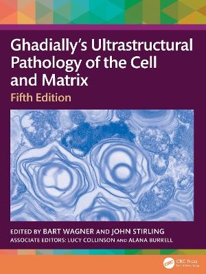
Ultrastructural Pathology of the Cell and Matrix
CRC Press (Verlag)
978-1-032-81189-5 (ISBN)
- Noch nicht erschienen (ca. April 2025)
- Versandkostenfrei innerhalb Deutschlands
- Auch auf Rechnung
- Verfügbarkeit in der Filiale vor Ort prüfen
- Artikel merken
Key Features:
Illustrates cell ultrastructure with electron micrographs
Reviews extracellular matrix structure
Assists pathologists in diagnosis of cellular pathologies
Includes contributions from an international team of leading researchers
Bart E. Wagner was born in The Hague, Holland, to Dutch parents in 1958 and moved to Sheffield, England, in 1960. He went to secondary school in Bakewell, Derbyshire. In 1976 he went to Portsmouth University in order to part study Marine Biology and came out with a BSc (Hons) in Biology. Fortunately, the degree was sufficiently general to enable him to take up a position as a trainee Biomedical Scientist in the Histopathology department in 1979 at the Royal Hallamshire Hospital NHS Trust in Sheffield. He passed the Institute of Biomedical Science (IBMS) Histopathology and Cytology Special Final in 1982, which for professional purposes is counted as equivalent to a MSc, and which made him a Fellow of the Institute of Biomedical Science. The project for the fellowship exam was a study of asteroid bodies in sarcoidosis which included histochemistry, immunohistochemistry and, where his interest in electron microscopy started, EM - all done whilst working in the main Histopathology laboratory. In 1984 he became the head of the electron microscopy section of the Histopathology Department at the Royal Hallamshire Hospital, sharing the hospital EM facilities with his counterparts at the Medical School, until renal pathology was moved to the Northern General Hospital in Sheffield in 1990. In 2012 he was moved back to the Royal Hallamshire Hospital where he continues to this day as head of the EM section. In 1984, there were EM facilities in 5 hospitals serving 8 departments in Sheffield. For technological reasons in diagnostic virology and tumour diagnosis, EM samples for those tests decreased to the extent that by 2005 rationalisation of diagnostic EM facilities in Sheffield had centralised to Bart. In addition, before improvements in molecular genetics (in approximately 2019) he received numerous paediatric liver samples from King’s College Hospital, London, and became the national centre for EM of skin for diagnosis of Ehlers-Danlos Syndrome. In 1997 Bart was awarded an IBMS travel fellowship to visit Professor Feroze N Ghadially in Ottawa, Canada, shortly after the 4th edition of Ultrastructural Pathology of the Cell and Matrix was published. Bart arrived in Prof Ghadially’s emeritus office with a large stack of electron micrograph photographic prints for his expert opinion. He was able to comment on most of the images - but not all - which spurred Bart into organising a 2-day national diagnostic EM conference in Sheffield in 1998 - followed a year later by the formal founding of the Association of Clinical Electron Microscopists (ACEM). He was the ACEM meeting secretary, organising annual meetings as a companion society to PathSoc until 2019. He is still on the executive of ACEM, but in the capacity of education and training secretary. He has been a TEM external quality assurance assessor for NEQAS based in Newcastle since 2018 to the present day. He has lectured on many diagnostic EM related subjects at numerous national and international meetings and on MSc courses. He has contributed EM to over 100 pathology publications. In 2008 he set up and completed an IBMS Diploma of Expert Practice in Ultrastructural Pathology - for which this updated book is essential reading. He is now an examiner for this qualification. Associate Professor John Stirling, College of Medicine and Public Health, Flinders University, Adelaide, South Australia. John has extensive experience in electron microscopy and cell ultrastructure gained during a career that spans almost sixty years. He started his career as an electron microscopist in 1965 in the Zoology Department of the University of Bristol (UK). After three years working with insect and animal tissues, he moved on to pursue a degree at Cardiff University (Wales), graduating with Honours in Zoology and Botany. As a postgraduate he then spent three years at the Tenovus Institute (Welsh National School of Medicine, Cardiff, Wales), specialising in breast cancer research, and subsequently publishing the first ultrastructural descriptions of the normal female mammary gland. John’s next move was to University College, Dublin (Republic of Ireland), as the inaugural manager of the Biological Sciences Electron Microscope Unit. In this role he oversaw the facility’s development and supported a range of biological research projects. While in Dublin, he played a key part in initiating the first Irish national electron microscopy conference, and was a founding member and Chairperson of the Irish Society of Electron Microscopy. In 1984 John moved to South Australia, becoming a diagnostic electron microscopist in Anatomical Pathology, principally at Flinders Medical Centre, Adelaide. Diagnostic ultrastructural pathology and immunogold labelling techniques were his specialties until he retired as the manager of the State’s Centre for Ultrastructural Pathology (SA Pathology, Adelaide). At Flinders Medical Centre, John became involved with Flinders University where he taught in biomedical courses, and was a member of the Medical Admissions Committee. John helped to establish the Bachelor of Medical Science, was a Topic Coordinator, and continues to contribute to the course. John’s other major professional involvement is with the Australian Institute of Medical and Clinical Science (AIMS). As a member of the AIMS South Australian/Northern Territory committee he has contributed to many local and national conferences as an organiser, presenter and workshop facilitator; he was also the co-editor of the Institute’s Journal, the Australian Journal of Medical Science. John received the Institute’s Gold Medal and was made an Honorary Life member in 2016. John’s publications cover immunogold labelling and a range of ultrastructural- and pathology-related topics. He has contributed chapters to a number of popular text books, including: Bancroft’s Theory and Practice of Histological Techniques (Elsevier); Laboratory Histopathology - A Complete Reference (Churchill Livingstone); and Antigen Retrieval Techniques: Immunohistochemistry and Molecular Morphology (Eaton Publishing). John also initiated, part-authored and was the Editor-in-Chief of Diagnostic electron microscopy: a practical guide to interpretation and technique (RMS/John Wiley). John has been a member of the Editorial Board of the Journal of Histochemistry and Cytochemistry, is an Associate Fellow of the Royal College of Pathologists of Australasia, a Fellow of the Royal Microscopical Society, and a member of the European Microscopy Society. John was the winner of the Specialist Area prize in the Inaugural South Australian Allied, Scientific and Complementary Health Excellence Awards in 2008. This was for his work on quality assurance for diagnostic electron microscopy, now established as an external quality assurance program run by the Royal College of Pathologists of Australasia (RCPA QAP). Dr Lucy Collinson is an electron microscopist with a background in microbiology and cell biology. She has a degree and PhD in Medical Microbiology and carried out post-doctoral research with Professor Colin Hopkins at the MRC Laboratory for Molecular Cell Biology (UCL) and Imperial College London, investigating membrane trafficking pathways in lysosome-related organelles in mammalian cells using light and electron microscopy as key techniques. She has run a series of biological electron microscopy facilities since 2004, at UCL and then at the Cancer Research UK London Research Institute, which became part of the new Francis Crick Institute in 2015. With a team of 10 electron microscopists and 3 physicists, she oversees more than 100 research projects with more than 60 research groups within the Francis Crick Institute, imaging across scales from proteins to whole organisms. Her microscopy and technology development interests include volume EM, correlative imaging techniques, cryo-microscopy, X-ray microscopy, image analysis, and microscope design and prototyping. Her group is using Citizen Science to gather hundreds of thousands of annotations of EM images to train deep machine learning algorithms to automatically recognise organelles in EM images through the Etch a Cell project on the Zooniverse platform. She is committed to open science through sharing of image data, protocols, software and hardware designs. She has co-authored more than 100 research and review papers, given more than 100 invited and keynote talks, and has sat on more than 45 international advisory boards, panels, and committees in advanced imaging. Dr Alana Burrell started her career by training in Veterinary Medicine at the Royal Veterinary College London (UK). After qualifying as a vet in 2015, she was accepted onto a joint PhD programme, split between the Royal Veterinary College London and Oxford Brookes University (UK). Her PhD work included the use of light and electron microscopy to characterise organelle diversity and dynamics for the Apicomplexan parasite Eimeria tenella. After completing her PhD, Alana moved to a position at the Francis Crick Institute in London (UK) within the Electron Microscopy Science Technology Platform (EM STP). In this role she has worked with a variety of biological samples (2D and 3D cell culture systems, bacterial and parasitic pathogens, animal tissue, and human tissue) and used both 2D and 3D advanced light and electron imaging techniques. She currently has a postdoctoral role, based within the EM STP at the Francis Crick Institute and as a visiting researcher at Imperial College London (UK), working on a large multimodal imaging project investigating antibody- mediated kidney transplant rejection.
Chapter 1. The Nucleus. Chapter 2. Centrosomes. Chapter 3. Mitochondria. Chapter 4. The Golgi Apparatus and Secretory Granules. Chapter 5. Endoplasmic reticulum. Chapter 6. Annulate lamellae - nucleopore complexes and tubulohelical membrane arrays. Chapter 7. Lysosomes. Chapter 8. Microbodies (peroxisomes and microperoxisomes). Chapter 9. Melanosomes. Chapter 10. Rod-shaped microtubulated bodies (Weibel-Palade bodies). Chapter 11. Intracytoplasmic Filaments. Chapter 12. Microtubules. Chapter 13. The cytosol (cytoplasmic matrix) and its inclusions. Chapter 14. Cell membrane and coat. Chapter 15. Cell junctions. Chapter 16. Cilia and flagella. Chapter 17. Extracellular matrix (extracellular components). Chapter 18. Exocytosis, endocytosis, cell-in-cell phenomena, cell surface structures, and glomerular podocytes (microvilli and foot processes). Chapter 19. The early development of electron microscope technology and techniques in biology and the biomedical sciences. Chapter 20. Current advances in Electron Microscopy technology and techniques with postulated applications in pathology.
| Erscheint lt. Verlag | 17.4.2025 |
|---|---|
| Zusatzinfo | 53 Tables, black and white; 8 Line drawings, black and white; 695 Halftones, black and white; 703 Illustrations, black and white |
| Verlagsort | London |
| Sprache | englisch |
| Maße | 210 x 280 mm |
| Themenwelt | Medizinische Fachgebiete ► Radiologie / Bildgebende Verfahren ► Radiologie |
| Studium ► 2. Studienabschnitt (Klinik) ► Pathologie | |
| Naturwissenschaften ► Biologie ► Zellbiologie | |
| ISBN-10 | 1-032-81189-7 / 1032811897 |
| ISBN-13 | 978-1-032-81189-5 / 9781032811895 |
| Zustand | Neuware |
| Informationen gemäß Produktsicherheitsverordnung (GPSR) | |
| Haben Sie eine Frage zum Produkt? |
aus dem Bereich


