Thieme Atlas of Anatomy
Thieme (Verlag)
978-1-68420-591-2 (ISBN)
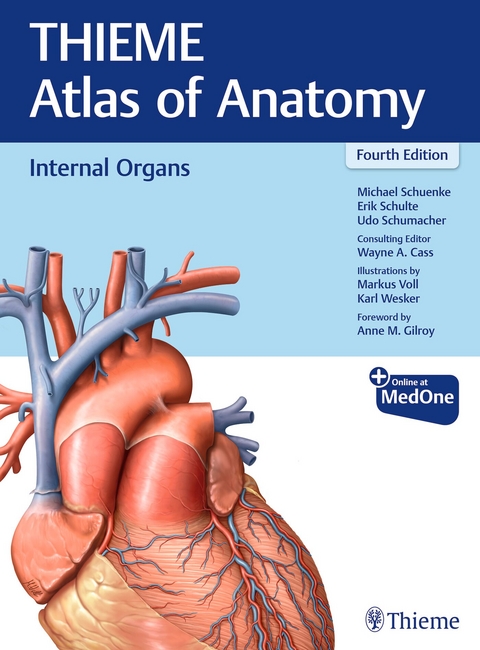
»Thieme Atlas of Anatomy: Internal Organs«, Fourth Edition, by renowned educators Michael Schuenke, Erik Schulte, and Udo Schumacher, along with consulting editor Wayne Cass, expands on prior editions. This volume features reorganized and updated sections, including additional coverage of clinical applications, radiologic procedures, and illustrations covering organs of the thorax, abdomen and pelvis, and perineum. As with the widely acclaimed prior editions, anatomic concepts are presented in a step-by-step sequence with system-by-system and topographical views.
The book lays a solid foundation of anatomic knowledge, with an introductory overview of each of the organs, including discussion of embryologic development. Each of the two regional units starts with a short overview chapter, followed by the structure and neurovasculature of the region and its organs. Subsequent chapters covering topographic regional anatomy support classroom learning and active dissection in the laboratory. The new edition further delineates anatomic relationships of inner organs, and the innervation and lymphatic systems of these organs.
Key Highlights:
- A total of 1,437 images, including new, exquisitely realistic illustrations by Markus Voll and Karl Wesker that reflect gender and ethnicity diversity
- A new chapter delineates cross-sectional thorax anatomy, with stunning images by vertebra level
- Organs of the Digestive System and their Neurovasculature features new diagnostic imaging for colorectal and small intestine tumors, as well as hepatic illustrations
- Salient points for each organ are summarized in 22 high-yield fact sheets
This atlas provides a robust resource for gross anatomy course instructors and medical and allied health students looking for a deeper dive into organ system anatomy. It also provides an outstanding reference for physical therapists, yoga instructors, and related professionals.
The THIEME Atlas of Anatomy series also includes two additional volumes, General Anatomy and Musculoskeletal System and Head, Neck, and Neuroanatomy.
All volumes of the THIEME Atlas of Anatomy series are available in softcover English/International Nomenclature and in hardcover with Latin nomenclature.
This print book includes complimentary access to a digital copy on https://medone.thieme.com.
Publisher's Note: Products purchased from Third Party sellers are not guaranteed by the publisher for quality, authenticity, or access to any online entitlements included with the product.
A Structure and Development of Organ Systems
1 Body Cavities
1.1 Definitions, Overview, and Evolution of Body Cavities
1.2 Organogenesis and the Development of Body Cavities
1.3 Compartmentalization of the Intraembryonic Coelom
1.4 Organization and Architecture of Body Cavities
2 Cardiovascular System
2.1 Overview and Basic Wall Structure
2.2 Terminal Vessels and Overview of the Major Blood Vessels
2.3 Cardiogenic Area, Development of the Heart Tube
2.4 Development of the Inner Chambers of the Heart and Fate of the Sinus Venosus
2.5 Cardiac Septation (Formation of Atrial, Interventricular, and Aorticopulmonary Septa)
2.6 Pre- and Postnatal Circulation and Common Congenital Heart Defects
3 Blood
3.1 Blood: Components
3.2 Blood: Cells
3.3 Blood: Bone Marrow
4 Lymphatic System
4.1 Overview of the Lymphatic System
4.2 Lymphatic Drainage Pathways
5 Respiratory System
5.1 Overview of the Respiratory System
5.2 Development of the Larynx, Trachea, and Lungs
5.3 Lung Development and Maturation
6 Digestive System
6.1 Overview of the Digestive System
6.2 Development and Differentiation of the Gastrointestinal Tract
6.3 Mesenteries and Primordia of the Digestive Organs in the Caudal Foregut Region; Stomach Rotation
6.4 Stomach Rotation and Organ Location in the Caudal Foregut Region; Formation of the Omental Bursa
6.5 Rotation of the Intestinal Loop and Development of Midgut and Hindgut Derivatives
6.6 Summary of the Development of the Midgut and Hindgut; Developmental Anomalies
7 Urinary System
7.1 Overview of the Urinary System
7.2 Development of the Kidneys, Renal Pelvis, and Ureters
7.3 Development of Nephrons, and the Urinary Bladder and Ureters; Developmental Anomalies
8 Genital System
8.1 Overview of the Genital System
8.2 Development of the Gonads
8.3 Development of the Genital Ducts
8.4 Comparison of Gender Differences and Relationship to the Urinary System
8.5 Comparison of Embryonic and Mature Structures
9 Endocrine System
9.1 Overview of the Endocrine System
9.2 Metabolism and Feedback Loop Regulation in the Endocrine System
10 Autonomic Nervous System
10.1 The Sympathetic and Parasympathetic Nervous Systems
10.2 Afferent Pathways of the Autonomic Nervous System and the Enteric Nervous System
10.3 Paraganglia
B Thorax
1 Overview and Diaphragm
1.1 Divisions of the Thoracic Cavity and Mediastinum
1.2 Diaphragm: Location and Projection onto the Trunk
1.3 Diaphragm: Structure and Main Openings
1.4 Diaphragm: Innervation, Blood, and Lymphatic Vessels
2 Overview of Neurovascular Structures
2.1 Arteries: Thoracic Aorta
2.2 Veins: Vena Cava and Azygos System
2.3 Lymphatic Vessels
2.4 Thoracic Lymph Nodes
2.5 Thoracic Innervation
3 Organs of the Cardiovascular System and their Neurovasculature
3.1 Location of the Heart in the Thorax
3.2 Pericardium: Location, Structure, and Innervation
3.3 Heart: Shape and Structure
3.4 Structure of the Heart Musculature (Myocardium)
3.5 Cardiac Chambers
3.6 Overview of the Cardiac Valves (Valve Plane and Cardiac Skeleton)
3.7 Cardiac Valves and Auscultation Sites
3.8 Radiographic Appearance of the Heart
3.9 Sonographic Appearance of the Heart: Echocardiography
3.10 Magnetic Resonance Imaging (MRI) of the Heart
3.11 Impulse Formation and Conduction System of the Heart; Electrocardiogram
3.12 Mechanical Action of the Heart
3.13 Coronary Arteries and Cardiac Veins: Classification and Topography
3.14 Coronary Arteries: Coronary Circulation
3.15 Coronary Heart Disease (CHD) and Heart Attack
3.16 Conventional Coronary Angiography (Heart Catheter Examination): Principle and Execution
3.17 Conventional Coronary Angiography: RAO and LAO Projections of the Coronary Arteries
3.18 Multislice Spiral Computed Tomography (MSCT) Coronary Angiography
3.19 Balloon Dilatation, Aortocoronary Venous and Arterial IMA Bypass
3.20 Lymphatic Drainage of the Heart
3.21 Innervation of the Heart
4 Organs of the Respiratory System and their Neurovasculature
4.1 Lungs: Location in the Thorax
4.2 Pleural Cavities
4.3 Boundaries of the Lungs and Parietal Pleura
4.4 Trachea
4.5 Lungs: Shape and Structure
4.6 Lungs: Segmentation
4.7 Functional Structure of the Bronchial Tree
4.8 Pulmonary Arteries and Veins
4.9 Bronchial Arteries and Veins
4.10 Functional Structure of the Vascular Tree
4.11 Innervation and Lymphatic Drainage of the Trachea, Bronchial Tree, and Lungs
4.12 Respiratory Mechanics
4.13 Radiological Anatomy of the Lungs and Vascular System
4.14 Computed Tomography of the Lungs
5 Esophagus and Thymus and their Neurovasculature
5.1 Esophagus: Location and Divisions
5.2 Esophagus: Inlet and Outlet, Opening and Closure
5.3 Esophagus: Wall Structure and Weaknesses
5.4 Arteries and Veins of the Esophagus
5.5 Lymphatic Drainage of the Esophagus
5.6 Innervation of the Esophagus
5.7 Thymus
6 Topographical Anatomy
6.1 Surface Anatomy, Topographical Regions, and Palpable Bony Landmarks
6.2 Anatomical Landmarks of the Thoracic Skeleton (Projection of Organs)
6.3 Structure of the Anterior Thoracic Wall and its Neurovascular Structures
6.4 Thoracic Organs in situ: Anterior, Lateral, and Inferior Views
6.5 Thoracic Organs in situ: Posterior Views
6.6 Heart: Pericardial Cavity
6.7 Overview of the Mediastinum
6.8 Posterior Mediastinum
6.9 Superior Mediastinum
6.10 Aortic Arch and Superior Thoracic Aperture
6.11 Clinical Aspects: Coarctation of the Aorta
6.12 Clinical Aspects: Aortic Aneurysm
7 Cross-Sectional Anatomy
7.1 Cross Sections through the Thorax at the Level of Vertebrae T1–T2
7.2 Cross Sections through the Thorax at the Level of Vertebrae T3–T4
7.3 Cross Sections through the Thorax at the Level of Vertebrae T5–T6
7.4 Cross Sections through the Thorax at the Level of Vertebrae T6–T7
7.5 Cross Sections through the Thorax at the Level of Vertebra T8
7.6 Cross Sections through the Thorax at the Level of Vertebrae T9–T10
7.7 Cross Sections through the Thorax at the Level of Vertebrae T10–T11
C Abdomen and Pelvis
1 Structure of the Abdominal and Pelvic Cavities: Overview
1.1 Architecture, Wall Structure, and Functional Aspects
1.2 Divisions of the Abdominal and Pelvic Cavities
1.3 Classification of Internal Organs Based on their Relationship to the Abdominal and Pelvic Cavities
2 Overview of Neurovascular Structures
2.1 Branches of the Abdominal Aorta: Overview and Paired Branches
2.2 Branches of the Abdominal Aorta: Unpaired and Indirect Paired Branches
2.3 Inferior Vena Caval System
2.4 Portal Venous System (Hepatic Portal Vein)
2.5 Venous Anastomoses in the Abdomen and Pelvis
2.6 Lymphatic Trunks and Lymph Nodes
2.7 Overview of the Lymphatic Drainage of Abdominal and Pelvic Organs
2.8 Autonomic Ganglia and Plexuses
2.9 Organization of the Sympathetic and Parasympathetic Nervous Systems
3 Organs of the Digestive System and their Neurovasculature
3.1 Stomach: Location, Shape, Divisions, and Interior View
3.2 Stomach: Wall Structure and Histology
3.3 Small Intestine: Duodenum
3.4 Small Intestine: Jejunum and Ileum
3.5 Large Intestine: Colon Segments
3.6 Large Intestine: Wall Structure, Cecum, and Vermiform Appendix
3.7 Large Intestine: Location, Shape, and Interior View of Rectum
3.8 Continence Organ: Structure and Components
3.9 Continence Organ: Function
3.10 Disorders of the Anal Canal: Hemorrhoidal Disease, Anal Abscesses, and Anal Fistulas
3.11 Colorectal Tumors: Frequency, Risk Factors, and Screening
3.12 Colorectal Tumors: Diagnostic Imaging and Surgical Treatment
3.13 Liver: Position and Relationship to Adjacent Organs
3.14 Liver: Peritoneal Relationships and Shape
3.15 Liver: Segmentation and Histology
3.16 Gallbladder and Bile Ducts: Location and Relationships to Adjacent Organs
3.17 Extrahepatic Bile Ducts and Pancreatic Ducts
3.18 Pancreas
3.19 Spleen
3.20 Branches of the Celiac Trunk: Arteries Supplying the Stomach, Liver, and Gallbladder
3.21 Branches of the Celiac Trunk: Arteries Supplying the Pancreas, Duodenum, and Spleen
3.22 Branches of the Superior Mesenteric Artery: Arteries Supplying the Pancreas, Small Intestine, and Large Intestine
3.23 Branches of the Inferior Mesenteric Artery: Arteries Supplying the Large Intestine
3.24 Branches of the Inferior Mesenteric Artery: Supply to the Rectum
3.25 Portal Vein: Venous Drainage of the Stomach, Duodenum, Pancreas, and Spleen
3.26 Superior and Inferior Mesenteric Veins: Venous Drainage of the Small and Large Intestine
3.27 Branches of the Inferior Mesenteric Vein: Venous Drainage of the Rectum
3.28 Lymphatic Drainage of the Stomach, Spleen, Pancreas, Duodenum, and Liver
3.29 Lymphatic Drainage of the Small and Large Intestine
3.30 Autonomic Innervation of the Liver, Gallbladder, Stomach, Duodenum, Pancreas, and Spleen
3.31 Autonomic Innervation of the Intestine: Distribution of the Superior Mesenteric Plexus
3.32 Autonomic Innervation of the Intestine: Distribution of the Inferior Mesenteric Plexus and Inferior Hypogastric Plexus
4 Organs of the Urinary System and their Neurovasculature
4.1 Overview of the Urinary Organs; the Kidneys in situ
4.2 Kidneys: Location, Shape, and Structure
4.3 Kidneys: Architecture and Microstructure
4.4 Renal Pelvis and Urinary Transport
4.5 Suprarenal Glands
4.6 Ureters in situ
4.7 Urinary Bladder in situ
4.8 Urinary Bladder, Bladder Neck, and Urethra: Wall Structure and Function
4.9 Functional Anatomy of Urinary Continence
4.10 Urethra
4.11 Arteries and Veins of the Kidneys and Suprarenal Glands: Overview
4.12 Arteries and Veins of the Kidneys and Suprarenal Glands: Variants
4.13 Lymphatic Drainage of the Kidneys, Suprarenal Glands, Ureter, and Urinary Bladder
4.14 Autonomic Innervation of the Urinary Organs and Suprarenal Glands
5 Organs of the Genital System and their Neurovasculature
5.1 Overview of the Genital Tract
5.2 Female Internal Genitalia: Overview
5.3 Female Internal Genitalia: Topographical Anatomy and Peritoneal Relationships; Shape and Structure
5.4 Female Internal Genitalia: Wall Structure and Function of the Uterus
5.5 Female Internal Genitalia: Positions of the Uterus and Vagina
5.6 Female Internal Genitalia: Epithelial Regions of the Uterus
5.7 Female Internal Genitalia: Cytologic Smear, Conization; Cervical Carcinoma
5.8 Female Internal Genitalia: Ovary and Follicular Maturation
5.9 Pregnancy and Childbirth
5.10 Male Genitalia: Accessory Sex Glands
5.11 Prostate Tumors: Prostatic Carcinoma, Prostatic Hyperplasia; Cancer Screening
5.12 Male Genitalia: Scrotum, Testis, and Epididymis
5.13 Male Genitalia: Seminiferous Structures and Ejaculate
5.14 Branches of the Internal Iliac Artery: Overview of Arteries Supplying the Pelvic Organs and Pelvic Wall
5.15 Vascularization of the Male Pelvic Organs
5.16 Vascularization of the Female Pelvic Organs
5.17 Vascularization of the Female Internal Genitalia and Urinary Bladder
5.18 Lymphatic Drainage of the Male and Female Genitalia
5.19 Autonomic Innervation of the Male Genitalia
5.20 Autonomic Innervation of the Female Genitalia
6 Topographical Anatomy
6.1 Surface Anatomy, Topographic Regions, and Palpable Bony Landmarks
6.2 Location of the Abdominal and Pelvic Organs and their Projection onto the Trunk Wall
6.3 Topography of the Opened Peritoneal Cavity (Supracolic Part and Infracolic Part)
6.4 Drainage Spaces and Recesses within the Peritoneal Cavity
6.5 Overview of the Mesenteries
6.6 Topography of the Omental Bursa
6.7 Topography of the Upper Abdominal Organs: Liver, Gallbladder, Duodenum, and Pancreas
6.8 Topography of the Upper Abdominal Organs: Stomach and Spleen
6.9 Cross-Sectional Anatomy of the Upper Abdominal Organs
6.10 Topography of the Small and Large Intestine
6.11 Diagnostic Imaging of the Small and Large Intestine: Plain-Film Abdominal Radiographs and Double-Contrast Studies
6.12 Diagnostic Imaging of the Small and Large Intestine: Intestinal Ultrasound, Computed Tomography, and MR Enterography
6.13 Topography of the Rectum
6.14 Retroperitoneum: Overview and Divisions
6.15 Retroperitoneum: Peritoneal Relationships
6.16 Retroperitoneum: Organs of the Retroperitoneum
6.17 Retroperitoneum: Location of the Kidneys
6.18 Peritoneal Relationships in the Anterior Abdominal Wall
6.19 Peritoneal Relationships in the Lesser Pelvis
6.20 Topography of Pelvic Connective Tissue, Levels of the Pelvic Cavity, and the Pelvic Floor
6.21 Suspensory Apparatus of the Uterus
6.22 Female Pelvis in situ
6.23 Male Pelvis in situ
6.24 Cross-Sectional Anatomy of the Female Pelvis
6.25 Cross-Sectional Anatomy of the Male Pelvis
D Neurovascular Supply to the Organs
1.1 Thymus
1.2 Esophagus
1.3 Heart
1.4 Pericardium
1.5 Lung and Trachea
1.6 Diaphragm
1.7 Liver, Gallbladder, and Spleen
1.8 Stomach
1.9 Duodenum and Pancreas
1.10 Jejunum and Ileum
1.11 Cecum, Vermiform Appendix, Ascending and Transverse Colon
1.12 Descending Colon and Sigmoid Colon
1.13 Rectum
1.14 Kidney, Ureter, and Suprarenal Gland
1.15 Urinary Bladder, Prostate, and Seminal Vesicle
1.16 Testis, Epididymis, and Ductus Deferens
1.17 Uterus, Uterine Tube, and Vagina
1.18 Uterine Tube and Ovary
E Organ Fact Sheets
1.1 Thymus
1.2 Pericardium
1.3 Heart
1.4 Trachea, Bronchi, and Lungs
1.5 Esophagus
1.6 Stomach
1.7 Small Intestine: Duodenum
1.8 Small Intestine: Jejunum and Ileum
1.9 Large Intestine: Cecum with Vermiform Appendix and Colon
1.10 Large Intestine: Rectum
1.11 Liver
1.12 Gallbladder and Bile Ducts
1.13 Pancreas
1.14 Spleen
1.15 Suprarenal Glands
1.16 Kidneys
1.17 Ureter
1.18 Urinary Bladder
1.19 Urethra
1.20 Vagina
1.21 Uterus and Uterine Tubes
1.22 Prostate and Seminal Vesicle
1.23 Epididymis and Ductus Deferens
1.24 Testis
1.25 Ovary
| Erscheinungsdatum | 20.11.2024 |
|---|---|
| Zusatzinfo | 1437 Illustrations |
| Verlagsort | New York |
| Sprache | englisch |
| Maße | 229 x 311 mm |
| Themenwelt | Studium ► 1. Studienabschnitt (Vorklinik) ► Anatomie / Neuroanatomie |
| ISBN-10 | 1-68420-591-3 / 1684205913 |
| ISBN-13 | 978-1-68420-591-2 / 9781684205912 |
| Zustand | Neuware |
| Haben Sie eine Frage zum Produkt? |
aus dem Bereich


