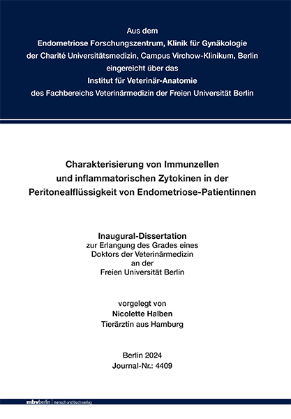
Charakterisierung von Immunzellen und inflammatorischen Zytokinen in der Peritonealflüssigkeit von Endometriose-Patientinnen
Seiten
2024
|
1. Auflage
Mensch & Buch (Verlag)
978-3-96729-235-0 (ISBN)
Mensch & Buch (Verlag)
978-3-96729-235-0 (ISBN)
Endometriose (EM) tritt Östrogen-abhängig auf und ist eine der häufigsten gynäkologischen Erkrankungen von Frauen im reproduktiven Alter. Das Auftreten von funktionalem Gewebe des Endometriums (Stroma und endometriale Drüsen) außerhalb der Uterushöhle, meist in Form von peritonealen Läsionen, definiert diese chronisch inflammatorische Erkrankung. Der Pathomechanismus ist ungeklärt und entsprechend sind keine kausal-therapeutischen Methoden etabliert. Es ist bekannt, dass verschiedene Komponenten des Immunsystems die Pathogenese der EM beeinflussen. Zudem zeigten Studien, dass neben endometrialem Gewebe auch Peritonealflüssigkeit immunkompetente Zellen beinhaltet. Ziel dieser Studie war es, die Rolle von Immunzellen und inflammatorischen Zytokinen in der Pathogenese und Etablierung von EM zu untersuchen. Hierzu wurden die Durchflusszytometrie, ein Bio-PlexTM Zytokin Assay und ein ELISA-Kit genutzt. Verschiedene Immunzellpopulationen und 27 Zytokine, Interleukine und Wachstumsfaktoren wurden in der Peritonealflüssigkeit von EM- und Kontroll-Patientinnen (benigne Geschehen wie Ovarialzysten oder Leiomyome) detektiert und charakterisiert. Zudem wurde die Konzentration des pro-inflammatorischen Neopterin, welches in verschiedenen chronischen Erkrankungen als Biomarker eingesetzt wird, analysiert. Die Ergebnisse zeigen, dass die Monozyten und Makrophagen die dominierenden Zellen in der Peritonealflüssigkeit der Frauen mit EM sind und die Zellgröße dieser Population positiv mit dem Schweregrad der EM korreliert. Zudem exprimieren sie HLA-DR signifikant geringer. Darüber hinaus sind signifikant erhöhte Konzentrationen an IL- 1β, IL-1Ra, IL-2, IL-4, IL-8, IL-10, IL-12 (p70), IL-17α, FGFb, G-CSF, MCP-1, MIP-1α, TNF-α und Neopterin in der Peritonealflüssigkeit der betroffenen Patientinnen zu finden. Die Konzentrationen von IL-12 (p70), IL-17α und FGFb sind speziell in milder EM signifikant hochreguliert, während IL-2, IL-8, MCP-1, TNF-α und MIP-1α in schwerer EM die höchsten Konzentrationen erreichen. Diese Resultate korrelieren mit der Annahme, dass veränderte Immunzellaktivitäten die Pathogenese der EM beeinflussen, indem sie die chronische Inflammation, Vaskularisation, Proliferation und Etablierung der EM-Läsionen fördern. Die geringere HLA-DR-Expression und veränderte Zellgröße der Makrophagen in EM zeigen, dass die Veränderungen qualitativer Art sind und diese Population eine entscheidende Rolle in der Pathogenese der EM spielen könnte. Die erhöhten Konzentrationen der pro- und antiinflammatorischen Zytokine weisen darauf hin, dass beide immunologischen Komponenten am komplexen Pathomechanismus der EM beteiligt sein könnten. Zudem wurde der Botenstoff Neopterin erstmalig auf diese Weise als neuartiger diagnostischer Marker in diesem detaillierten Gesamtkontext analytisch betrachtet und muss auch in künftigen Studien als potentieller Marker im Rahmen der EM bedacht werden. "Characterization of immune cells and inflammatory cytokines in the peritoneal fluid of patients with endometriosis"
Endometriosis (EM) is an estrogen-dependent disease and one of the most common benign gynecological disorders of women in reproductive age. The appearance of functional endometrial tissue (stroma and endometrial glands) outside the uterine cavity, mostly as small peritoneal lesions, defines this chronic inflammatory disease. The pathomechanism remains unclear, thus there are no causal therapeutic methods established so far. It is known that different components of the immune system influence the pathogenesis of EM. Furthermore, studies have shown that besides endometriotic tissue, peritoneal fluid also generally contains immunocompetent cells. The aim of this study was to evaluate the potential role of immune cells and their inflammatory cytokines in the pathogenesis and establishment of EM. For this, flow cytometry, a Bio-PlexTM Cytokine Assay and an ELISA-Kit have been used. Different immune cell populations and 27 inflammatory cytokines, interleukins, and growth factors in the peritoneal fluid of EM patients and control patients (benign disorders like ovarian cysts or fibroids) have been detected and characterized. Additionally, the concentration of pro-inflammatory Neopterin, which is used as a biomarker in several chronic diseases, was analyzed. The results show that monocytes and macrophages are the dominating cells in the peritoneal fluid of women with EM and there is a positive correlation of cell size and severity of EM regarding this population. Additionally, their expression of HLA-DR is significantly lower. Furthermore, there are significantly higher levels of IL-1β, IL-1Ra, IL-2, IL-4, IL-8, IL-10, IL-12 (p70), IL-17α, FGFb, G-CSF, MCP-1, MIP-1α, TNF-α and Neopterin in the peritoneal fluid of affected patients. Especially the concentrations of IL-12 (p70), IL-17α and FGFb are significantly upregulated in mild EM, while IL-2, IL-8, MCP-1, TNF-α and MIP-1α show the highest levels in women with severe EM. These findings correlate with the assumption that altered immune cell activities influence the pathogenesis of EM by promoting chronic inflammation, vascularization, proliferation, and maintenance of endometriotic lesions. Lower expression of HLA-DR and varying cell size of monocytes and macrophages in women with EM show that there are qualitative alterations, and this population seems to play a crucial role in the pathogenesis of EM. The higher concentrations of pro- and anti-inflammatory cytokines indicate that both components of the immune response seem to take part in the complex pathomechanism of EM. Furthermore, it was the first time that Neopterin was analyzed as a potential new biomarker in this special and detailed context and this messenger has to be considered as a diagnostic parameter for EM in future studies.
Endometriosis (EM) is an estrogen-dependent disease and one of the most common benign gynecological disorders of women in reproductive age. The appearance of functional endometrial tissue (stroma and endometrial glands) outside the uterine cavity, mostly as small peritoneal lesions, defines this chronic inflammatory disease. The pathomechanism remains unclear, thus there are no causal therapeutic methods established so far. It is known that different components of the immune system influence the pathogenesis of EM. Furthermore, studies have shown that besides endometriotic tissue, peritoneal fluid also generally contains immunocompetent cells. The aim of this study was to evaluate the potential role of immune cells and their inflammatory cytokines in the pathogenesis and establishment of EM. For this, flow cytometry, a Bio-PlexTM Cytokine Assay and an ELISA-Kit have been used. Different immune cell populations and 27 inflammatory cytokines, interleukins, and growth factors in the peritoneal fluid of EM patients and control patients (benign disorders like ovarian cysts or fibroids) have been detected and characterized. Additionally, the concentration of pro-inflammatory Neopterin, which is used as a biomarker in several chronic diseases, was analyzed. The results show that monocytes and macrophages are the dominating cells in the peritoneal fluid of women with EM and there is a positive correlation of cell size and severity of EM regarding this population. Additionally, their expression of HLA-DR is significantly lower. Furthermore, there are significantly higher levels of IL-1β, IL-1Ra, IL-2, IL-4, IL-8, IL-10, IL-12 (p70), IL-17α, FGFb, G-CSF, MCP-1, MIP-1α, TNF-α and Neopterin in the peritoneal fluid of affected patients. Especially the concentrations of IL-12 (p70), IL-17α and FGFb are significantly upregulated in mild EM, while IL-2, IL-8, MCP-1, TNF-α and MIP-1α show the highest levels in women with severe EM. These findings correlate with the assumption that altered immune cell activities influence the pathogenesis of EM by promoting chronic inflammation, vascularization, proliferation, and maintenance of endometriotic lesions. Lower expression of HLA-DR and varying cell size of monocytes and macrophages in women with EM show that there are qualitative alterations, and this population seems to play a crucial role in the pathogenesis of EM. The higher concentrations of pro- and anti-inflammatory cytokines indicate that both components of the immune response seem to take part in the complex pathomechanism of EM. Furthermore, it was the first time that Neopterin was analyzed as a potential new biomarker in this special and detailed context and this messenger has to be considered as a diagnostic parameter for EM in future studies.
| Erscheinungsdatum | 30.05.2024 |
|---|---|
| Verlagsort | Berlin |
| Sprache | deutsch |
| Maße | 148 x 210 mm |
| Gewicht | 260 g |
| Themenwelt | Medizin / Pharmazie ► Medizinische Fachgebiete ► Gynäkologie / Geburtshilfe |
| Veterinärmedizin ► Vorklinik | |
| Schlagworte | Bauchfell • cytokines • Endometriose • Endometriose-Patientinnen • Endometriosis • Entzündung • Immunzellen • inflammation • inflammatorische Zytokinen • Interleukine • interleukins • Peritonealflüssigkeit • Peritoneum • Zytokine |
| ISBN-10 | 3-96729-235-5 / 3967292355 |
| ISBN-13 | 978-3-96729-235-0 / 9783967292350 |
| Zustand | Neuware |
| Informationen gemäß Produktsicherheitsverordnung (GPSR) | |
| Haben Sie eine Frage zum Produkt? |
Mehr entdecken
aus dem Bereich
aus dem Bereich
Buch | Hardcover (2024)
Urban & Fischer in Elsevier (Verlag)
94,00 €
alles für Station, Praxis und Facharztprüfung
Buch (2023)
Thieme (Verlag)
165,00 €


