Teaching Atlas of Pediatric Imaging
Seiten
2006
Thieme Medical Publishers Inc (Verlag)
978-1-58890-339-6 (ISBN)
Thieme Medical Publishers Inc (Verlag)
978-1-58890-339-6 (ISBN)
125 cases addressing "real-life" clinical problems
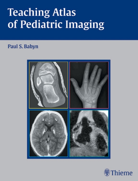
Complete with the insights of leading pediatric radiologists, Teaching Atlas of Pediatric Imaging provides 125 cases that address the challenging "real-life" clinical problems that you are likely to encounter. Each chapter presents a different case with a complete patient work-up that includes clinical presentation, diagnosis, differential diagnoses, radiological and clinical findings, treatment summary and suggested readings. With a view to providing the opportunity for self-assessment, the authors omit the diagnosis from the first pages of each case to enable self-testing and review.
Highlights:
Easy-to-access arrangement of cases based on anatomy: head and neck, chest, heart, abdomen, pelvis, and the musculoskeletal system
Coverage of a wide spectrum of diseases, from the very common to more important uncommon entities, including congenital heart disease, bone dysplasias and more
Differential diagnoses for each case, as well as information on etiology, pathology, treatment, and complications
"Pearls" and "Pitfalls" that help you identify important points and avoid errors in image interpretation
Here is a valuable resource for the clinician at every level, from the resident preparing for the radiology board examinations, to the practitioner seeking the Certificate of Added Qualification in Pediatric Radiology, to the general radiologist or pediatrician seeking a practical reference text.
Associate Professor, Department of Medical Imaging, University of Toronto & Radiologist in Chief, Department of Diagnostic Imaging, Hospital for Sick Children, Toronto, ON, Canada
| Erscheint lt. Verlag | 18.4.2006 |
|---|---|
| Zusatzinfo | 823 Illustrations |
| Verlagsort | New York |
| Sprache | englisch |
| Maße | 216 x 279 mm |
| Gewicht | 2057 g |
| Themenwelt | Medizin / Pharmazie ► Medizinische Fachgebiete ► Pädiatrie |
| Medizinische Fachgebiete ► Radiologie / Bildgebende Verfahren ► Nuklearmedizin | |
| Medizinische Fachgebiete ► Radiologie / Bildgebende Verfahren ► Radiologie | |
| ISBN-10 | 1-58890-339-7 / 1588903397 |
| ISBN-13 | 978-1-58890-339-6 / 9781588903396 |
| Zustand | Neuware |
| Haben Sie eine Frage zum Produkt? |
Mehr entdecken
aus dem Bereich
aus dem Bereich
Buch | Softcover (2022)
Lehmanns Media (Verlag)
39,95 €
Lehrbuch für Breast Care Nurses und Fachpersonen in der Onkologie
Buch | Hardcover (2020)
Hogrefe (Verlag)
50,00 €


