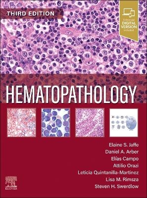
Hematopathology
Elsevier - Health Sciences Division (Verlag)
978-0-323-83165-9 (ISBN)
Helps you navigate the latest changes in the classification of hematolymphoid neoplasms, providing guidance for use of both the International Consensus Classification (ICC) and 5th edition of the WHO classification.
Incorporates the latest molecular/cytogenetic information, regarding newly recognized entities and the latest diagnostic criteria.
Provides you with today's most effective guidance in evaluating specimens from the lymph nodes, bone marrow, peripheral blood, and more, with authoritative information on the pathogenesis, clinical and pathologic diagnosis, and treatment for each.
Details the latest insights on the molecular biology of benign and malignant hematologic disorders.
Features more than 1,100 high-quality color images that mirror the findings you encounter in practice.
Uses an easy-to-navigate, templated format with standard headings in each chapter.
Includes information on disease progression and prognosis, helping you better understand the clinical implications of diagnosis.
Shares the knowledge and expertise of new editors, Drs. Lisa Rimsza, Attilio Orazi, and Steven Swerdlow, providing expertise in molecular diagnostics, bone marrow and lymph node biopsies.
An eBook version is included with purchase. The eBook allows you to access all of the text, figures and references, with the ability to search, customize your content, make notes and highlights, and have content read aloud. Additional digital ancillary content may publish up to 6 weeks following the publication date.
Dr. Elaine Jaffe is regarded by her peers as one of the most pre-eminent hematopathologists of her generation. She is most widely known for her work regarding the pathophysiology and prognosis of malignant lymphomas, as well as her unparalleled work to understand how they respond to treatment. Dr. Jaffe led the effort to develop the World Health Organization classification of tumors of the hematopoietic and lymphoid tissues published in 2001, a classification that rapidly became the international standard. Dr. Daniel Arber is an internationally recognized expert in the diagnosis and classification of hematopoietic tumors, including malignant lymphoma, acute and chronic leukemia, myeloproliferative disorders, myelodysplastic syndrome and tumors of the spleen. the clinical director of the Biomedical Diagnostic Centre of Hospital Clinic and a full Professor of Anatomical Pathology at the University of Barcelona, where he also teaches in the Department of Anatomical Pathology, Pharmacy and Microbiology. He is the Spanish coordinator of the International Cancer Genome Consortium. Professor Dr. Leticia Quintanilla de Fend, head of the Core Facility and Senior Physician at the Institute of Pathology has extensive experience in the interpretation of mouse models. From 2000 until 2008 she was a director of the mouse pathology at the Institute of Pathology of the Helmholtz Center Munich and the Pathology Screen of the German Mouse Clinic (GMC) and established various special procedures for the analysis of mouse models. Professor Quintanilla de Fend is involved in the establishment of standards in the pathology of mouse models within EU networks. Dr. Attilio Orazi is an internationally renowned academic hematopathologist whose diagnostic expertise spans all areas of hematopathology. However, he is best known for his expertise and scholarly accomplishments as a bone marrow pathologist. He is a member of the Scientific Committee of the European Bone Marrow Working Group, has directed courses concerning the assessment of bone marrow disorders and splenic pathology at the United States and Canadian Academy of Pathology and was the co-organizer of the 2007 Workshop of the Society of Hematopathology.
PART I Technical Aspects PART II Normal and Reactive Conditions of Hematopoietic Tissues SECTION 1 . MATURE B-CELL NEOPLASMS SECTION 2 . MATURE T-CELL AND NK-CELL NEOPLASMS SECTION 3 . PRECURSOR B- AND T-CELL NEOPLASMS PART IV Myeloid, Histiocytic, and Related Proliferations PART V Immunodeficiency Disorders PART VI Site-Specific Issues in the Diagnosis of Lymphoma and Leukemia
| Erscheinungsdatum | 03.09.2024 |
|---|---|
| Zusatzinfo | Approx. 2260 illustrations (2200 in full color); Illustrations |
| Verlagsort | Philadelphia |
| Sprache | englisch |
| Maße | 216 x 276 mm |
| Gewicht | 3900 g |
| Themenwelt | Studium ► 2. Studienabschnitt (Klinik) ► Pathologie |
| ISBN-10 | 0-323-83165-6 / 0323831656 |
| ISBN-13 | 978-0-323-83165-9 / 9780323831659 |
| Zustand | Neuware |
| Informationen gemäß Produktsicherheitsverordnung (GPSR) | |
| Haben Sie eine Frage zum Produkt? |
aus dem Bereich


