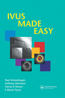
IVUS Made Easy
Seiten
2005
Informa Healthcare (Verlag)
978-1-84184-595-1 (ISBN)
Informa Healthcare (Verlag)
978-1-84184-595-1 (ISBN)
- Titel erscheint in neuer Auflage
- Artikel merken
Zu diesem Artikel existiert eine Nachauflage
Intravascular ultrasound (IVUS) is a tomographic imaging modality performed during coronary angiography that allows the simultaneous assessment of lumen, vessel wall, and atherosclerotic plaque. IVUS has been established as the method of choice for the detection and serial observation of transplant vasculopathy, and more recently, for the serial observation of atherosclerotic plaque burden in atherosclerosis progression/regression trials. When performed by an operator familiar with interventional, percutaneous techniques, the rate of complications during IVUS imaging is exceedingly low.
IVUS Made Easy provides an introduction to coronary imaging with intravascular ultrasound (IVUS). It consists of a brief, practical text and corresponding illustrated IVUS images. The format is uniform throughout, with a non-illustrated IVUS image displayed together with an illustrated copy. Based on the previously published An Atlas and Manual of Coronary Intravascular Ultrasound Imaging, this guide provides expanded descriptions of the practical aspects of IVUS and includes additional information in short case presentations.
The book begins with the principle of IVUS imaging. It discusses normal arterial anatomy by IVUS, image artifacts and IVUS measurements. It then discusses plaque (atheroma) morphology and clinical applications.
Case studies include:
Coronary arteritis
Angiographically indeterminate lesion
Indeterminate lesion: plaque rupture
Intedeterminate lesion after angioplasty
Intracoronary thrombus (2 cases)
Complications of IVUS: dissection
Chronic coronary arterial wall dissection behind stent
Intramural hematoma post-PCI
Serial IVUS: regression
Cleveland Clinic Foundation, Cleveland, USA Cardiology Associates PSC, Edgewood KY, USA The Cleveland Clinic Foundation, Cleveland, USA
1. Principle of IVUS Imaging 2. Normal Arterial Anatomy by IVUS 3. Image Artifacts 4. IVUS Measurements 5. Plaque Morphology 6. Clinical Applications 7. Conclusions 8. References
| Erscheint lt. Verlag | 13.9.2005 |
|---|---|
| Zusatzinfo | 95 Halftones, color; 20 Halftones, black and white |
| Sprache | englisch |
| Maße | 156 x 234 mm |
| Gewicht | 204 g |
| Themenwelt | Medizinische Fachgebiete ► Innere Medizin ► Kardiologie / Angiologie |
| Medizinische Fachgebiete ► Radiologie / Bildgebende Verfahren ► Sonographie / Echokardiographie | |
| ISBN-10 | 1-84184-595-7 / 1841845957 |
| ISBN-13 | 978-1-84184-595-1 / 9781841845951 |
| Zustand | Neuware |
| Informationen gemäß Produktsicherheitsverordnung (GPSR) | |
| Haben Sie eine Frage zum Produkt? |
Mehr entdecken
aus dem Bereich
aus dem Bereich
in Fällen, Fragen und Antworten
Buch | Softcover (2024)
Urban & Fischer in Elsevier (Verlag)
89,00 €
Diagnostik und interventionelle Therapie | 2 Bände
Buch (2024)
Deutscher Ärzteverlag
349,99 €


