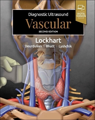
Diagnostic Ultrasound: Vascular
Churchill Livingstone (Verlag)
978-0-443-28366-6 (ISBN)
Provides a wide range of anatomic detail, technical factors, and diagnostic criteria to guide accurate application of ultrasound throughout the body
Covers new and evolving techniques such as the increasing use of microbubble imaging to enhance image resolution, distinguish vessels more clearly, and minimize noise and background signals
Details the latest information across several ACR RADS criteria, and contains extensive new material from the LI-RADS, GB-RADS, and transplant criteria, which now include Doppler ultrasound with its noninvasive methodology rated highly for appropriate use
Reflects an increased use of Doppler extremity evaluations due to ongoing COVID-19 diagnoses and a higher incidence of venous thrombosis
Contains updated ACR Appropriateness Criteria regarding the new “highly appropriate” ratings, as well as new Intersocietal Accreditation Commission (IAC) recommendations in numerous diagnosis chapters
Contains a gallery of typical and atypical ultrasound appearances covering a wide spectrum of disease, correlated with CT and MR imaging where appropriate, and detailed artistic renderings
Features image-rich chapters on vascular ultrasound techniques, covering grayscale, color, power, and spectral (pulsed) Doppler imaging, as well as imaging artifacts
Contains time-saving reference features such as succinct and bulleted text, a variety of test data tables, a Key Facts section that begins in each chapter, annotated images, and an extensive index
An ideal reference for radiologists, sonographers, vascular surgeons, and those who are training in these fields
Includes an eBook version that enables you to access all text, figures, and references, with the ability to search, customize your content, make notes and highlights, and have content read aloud
Mark E. Lockhart, MD, MPH, is Professor of Radiology at the Department of Radiology in the School of Medicine at the University of Alabama at Birmingham in Birmingham, Alabama
Part I: TechniqueSection 1: ModalitiesColor Doppler
Power Doppler
Spectral Doppler: General Waveform Concepts
Contrast-Enhanced Ultrasound: Basic Technique in Liver and Kidney
Volumetric Blood Flow
Microvascular ImagingSection 2: ArtifactsAliasing, Blooming, and Twinkling ArtifactPart II: AnatomySection 1: Head and NeckNeck Overview
Cervical Carotid Arteries
Vertebral Arteries
Neck VeinsSection 2: Chest and AbdomenThoracic Outlet
Ribs and Intercostal Space
Liver
Spleen
Pancreas
Kidneys
Aorta and Inferior Vena Cava
Mesenteric Vessels
Iliac Arteries and Veins
Penile
Scrotum
Gynecologic StructuresSection 3: ExtremitiesUpper Limb
Arm Overview
Arm Vessels
Forearm Overview
Wrist Overview
Hand Overview
Wrist and Hand Vessels
Lower Limb
Groin Overview
Femoral Vessels and Nerves
Lower Leg Overview
Lower Leg Vessels
Foot VesselsPart III: DiagnosesSection 1: Head and NeckParotid Vascular Lesion
Venous Vascular Malformation
Jugular Vein Thrombosis
Carotid Artery Dissection in Neck
Carotid Stenosis/Occlusion
Carotid-Jugular Fistula
Vertebral Stenosis/Occlusion
Atypical Carotid Waveforms
Arteritis/Takayasu
IABP and Other DevicesSection 2: Chest and AbdomenAorta and Vena CavaAortic/Iliac Aneurysm
Aortoiliac Occlusive Disease
IVC Obstruction
Aortic Dissection
Aortic Stent Endoleak
Celiac Artery Stenosis
SMA Stenosis
Median Arcuate Ligament Syndrome
Liver
Portal Hypertension
Approach to Hepatic Sonography
Portosystemic Collaterals
Transjugular Intrahepatic Portosystemic Shunt (TIPS)
Portal Vein Occlusion
Budd-Chiari Syndrome
Portal Vein Gas
Hepatic Tumor Thrombus/Tumor-in-Vein
Spleen
Splenic Vascular Disorders
Splenic Artery Aneurysm
Splenic Vein Thrombosis
Kidney
Renal Artery Stenosis
Renal Vein Thrombosis
Renal Infarct
Perinephric Hematoma
Tumor Vascularity
Renal AVF/AVM
Renal Vein Nutcracker
High Resistive Index
Reproductive
Varicocele
Testicular Torsion/Infarct
Penile
Ovarian Torsion
Uterine AVM/PSA
Uterine Fibroid Embolization
Pelvic CongestionSection 3: TransplantsKidney
Approach to Sonography of Renal Allografts
Transplant Renal Artery Stenosis
Transplant Renal Artery Thrombosis
Transplant Renal Vein Thrombosis
Renal Transplant Arteriovenous Fistula
Renal Transplant Pseudoaneurysm
Renal Transplant Rejection
Delayed Renal Graft Function
Liver
Liver Transplant Hepatic Artery Stenosis/Thrombosis
Liver Transplant Portal Vein Stenosis/Thrombosis
Liver Transplant Hepatic Venous Stenosis/Thrombosis
Liver Transplant Biliary Stricture
Hepatic Artery Aneurysm/Pseudoaneurysm
Pancreas
Pancreas Transplant: Normal vs. RejectionSection 4: ExtremitiesArteries
Peripheral Arterial Occlusive Disease
Peripheral Arterial Pseudoaneurysm
Peripheral Arteriovenous Fistula
Peripheral Artery Bypass
Wrist Modified Allen Test
Hemodialysis Stenosis/Occlusion
Hemodialysis Failure to Mature
Hemodialysis Steal
Veins
Deep Vein Thrombosis
Varicose Veins/Incompetent Perforator
Iliac and IVC Venous Occlusion
Soft Tissues
Limb Hemangioma
Vascular Dilatation or Inflammation
Vascular Anomaly
Vascular Tumor/LeiomyosarcomaSection 5: InterventionsThrombin PSA Repair
Aneurysm Coiling
| Erscheinungsdatum | 22.08.2024 |
|---|---|
| Zusatzinfo | Approx. 1880 full-color images and illustrations; Illustrations |
| Verlagsort | London |
| Sprache | englisch |
| Maße | 216 x 276 mm |
| Gewicht | 2130 g |
| Themenwelt | Medizinische Fachgebiete ► Innere Medizin ► Kardiologie / Angiologie |
| Medizinische Fachgebiete ► Radiologie / Bildgebende Verfahren ► Radiologie | |
| Medizinische Fachgebiete ► Radiologie / Bildgebende Verfahren ► Sonographie / Echokardiographie | |
| ISBN-10 | 0-443-28366-4 / 0443283664 |
| ISBN-13 | 978-0-443-28366-6 / 9780443283666 |
| Zustand | Neuware |
| Haben Sie eine Frage zum Produkt? |
aus dem Bereich


