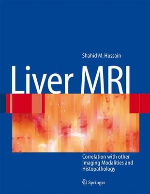
Liver MRI
Springer Berlin (Verlag)
978-3-540-25552-9 (ISBN)
- Titel erscheint in neuer Auflage
- Artikel merken
In March 1973, a short paper was published in Nature, entitled „Image formation by induced local int- action; examples employing nuclear magnetic resonance. “ The author was Paul C. Lauterbur, a Prof- sor of Chemistry at the State University of New York at Stony Brook. In this seminal manuscript the - thor described a new imaging technique which moved the single dimension of NMR spectroscopy to the dual dimension of spatial orientation, thereby resulting in the foundation of modern magnetic re- nance (MR) imaging. Over the ensuing years, MR imaging has assumed an increasingly important role in clinical imaging. It distinguishes itself from other imaging modalities, such as ultrasound (US) or computed tomography (CT), by the unique ability to visualize specific tissue components in a non-- vasive manner. In the earlier days, diagnostic MR imaging was limited to cerebral and musculoskeletal diseases. - aging of other areas which are more prone to movement through breathing (abdominal) or pulsation motions (cardiac) became available more recently, with the introduction of faster sequences and the - velopment of more dedicated MR imaging coils. Currently, liver MRI is considered the cornerstone of abdominal imaging. In most centers, however, liver MRI is still employed largely as a problem solving modality, when US and CT have provided an unsatisfactory outcome.
High-Fluid Content Liver Lesions.- Abscesses — Pyogenic Type.- Biliary Hamartomas (von Meyenberg Complexes).- Cyst I — Typical Small.- Cyst II — Typical Large with MR-CT Correlation.- Cyst III — Multiple Small Lesions with MR-CT-US Comparison.- Cyst IV — Adult Polycystic Liver Disease.- Cystadenoma / Cystadenocarcinoma.- Hemangioma I — Typical Small.- Hemangioma II — Typical Medium-Sized with Description of Pathology.- Hemangioma III — Typical Giant.- Hemangioma IV — Giant Type with a Large Central Scar.- Hemangioma V — Atypical, Flash-Filling with Perilesional Enhancement.- Hemangioma VI — Multiple with Perilesional Enhancement.- Hemorrhage.- Hemorrhage — Within a Solid Tumor.- Mucinous Metastasis — Mimicking an Hemangioma.- Solid Liver Lesions.- Colorectal Metastases I — Typical Lesion.- Colorectal Metastases II — Typical Multiple Lesions.- Colorectal Metastases III — Metastasis Versus Cyst.- Colorectal Metastases IV — Metastasis Versus Hemangiomas.- Liver Metastases V — Large, Mucinous, Mimicking a Primary Liver Lesion.- Colorectal Metastases VI — with Portal Vein and Bile Duct Encasement.- Colorectal Metastases VII — Recurrent Disease Versus RFA Defect.- Breast Carcinoma Liver Metastases.- Kahler’s Disease (Multiple Myeloma) Liver Metastases.- Melanoma Liver Metastases I — Focal Type.- Melanoma Liver Metastases II — Diffuse Type.- Neuroendocrine Tumor I — Typical Liver Metastases.- Neuroendocrine Tumor II — Pancreas Tumor Metastases.- Neuroendocrine Tumor III — Gastrinoma Liver Metastases.- Neuroendocrine Tumor IV — Carcinoid Tumor Liver Metastases.- Neuroendocrine Tumor V — Peritoneal Spread.- Ovarian Tumor Liver Metastases — Mimicking Giant Hemangioma.- Renal Cell Carcinoma Liver Metastasis.- Cirrhosis I — Liver Morphology.- Cirrhosis II — Regenerative Nodules and Confluent Fibrosis.- Cirrhosis III — Dysplastic Nodules.- Cirrhosis IV — Dysplastic Nodules — HCC Transition.- Cirrhosis V — Cyst in a Cirrhotic Liver.- Cirrhosis VI — Multiple Cysts in a Cirrhotic Liver.- Cirrhosis VII — Hemangioma in a Cirrhotic Liver.- HCC in Cirrhosis I — Typical Small with Pathologic Correlation.- HCC in Cirrhosis II — Small With and Without a Tumor Capsule.- HCC in Cirrhosis III — Nodule-in-Nodule Appearance.- HCC in Cirrhosis IV — Mosaic Pattern with Pathologic Correlation.- HCC in Cirrhosis V — Typical Large with Mosaic and Capsule.- HCC in Cirrhosis VI — Mosaic Pattern with Fatty Infiltration.- HCC in Cirrhosis VII — Large Growing Lesion with Portal Invasion.- HCC in Cirrhosis VIII — Segmental Diffuse with Portal Vein Thrombosis.- HCC in Cirrhosis IX — Multiple Lesions Growing on Follow-up.- HCC in Cirrhosis X — Capsular Retraction and Suspected Diaphragm Invasion.- HCC in Cirrhosis XI — Diffuse Within the Entire Liver with Portal Vein Thrombosis.- HCC in Cirrhosis XII — With Intrahepatic Bile Duct Dilatation.- Focal Nodular Hyperplasia I — Typical with Large Central Scar and Septa.- Focal Nodular Hyperplasia II — Typical with Pathologic Correlation.- Focal Nodular Hyperplasia III — Typical with Follow-up Examination.- Focal Nodular Hyperplasia IV — Multiple FNH Syndrome.- Focal Nodular Hyperplasia V — Fatty FNH with Concurrent Fatty Adenoma.- Focal Nodular Hyperplasia VI — Atypical with T2 Dark Central Scar.- Hepatic Angiomyolipoma — MR-CT Comparison.- Hepatic Lipoma — MR-CT-US Comparison.- Hepatocellular Adenoma I — Typical with Pathologic Correlation.- Hepatocellular Adenoma II — Large Exophytic with Pathologic Correlation.- Hepatocellular Adenoma III — Typical Fat-Containing.- Hepatocellular Adenoma IV — With Large Hemorrhage.- Hepatocellular Adenoma V — Multiple in Fatty Liver (Non-OC-Dependent).- Hepatocellular Adenoma VI — Multiple in Fatty Liver (OC-Dependent).- HCC in Non-Cirrhotic Liver I — Small with MR-Pathologic Correlation.- HCC in Non-Cirrhotic Liver II — Large with MR-Pathologic Correlation.- HCC in Non-Cirrhotic Liver III — Large Lesion with Inconclusive CT.- HCC in Non-Cirrhotic Liver IV — Cholangiocellular or Combined Type.- HCC in Non-Cirrhotic Liver V — Central Scar and Capsule Rupture.- HCC in Non-Cirrhotic Liver VI — Capsule with Pathologic Correlation.- HCC in Non-Cirrhotic Liver VII — Very Large with Pathologic Correlation.- HCC in Non-Cirrhotic Liver VIII — Vascular Invasion and Satellite Nodules.- HCC in Non-Cirrhotic Liver IX — Adenoma-Like HCC with Pathologic Correlation.- Intrahepatic Cholangiocarcinoma — With Pathologic Correlation.- Telangiectatic Hepatocellular Lesion.- Diffuse (Depositional) Liver Diseases.- Focal Fatty Infiltration Mimicking Metastases.- Focal Fatty Sparing Mimicking Liver Lesions.- Hemosiderosis — Iron Deposition, Acquired Type.- Hemochromatosis — Severe Type.- Hemochromatosis with Solitary HCC.- Hemochromatosis with Multiple HCC.- Thalassemia with Iron Deposition.- Vascular Liver Lesions.- Arterioportal Shunt I — Early Enhancing Lesion in a Cirrhotic Liver.- Arterioportal Shunt II — Early Enhancing Lesion in a Non-Cirrhotic Liver.- Budd-Chiari Syndrome I — Abnormal Enhancement and Intrahepatic Collaterals.- Budd-Chiari Syndrome II — Gradual Deformation of the Liver.- Budd-Chiari Syndrome III — Nodules Mimicking Malignancy.- Hereditary Hemorrhagic Telangiectasia or Rendu-Osler-Weber Disease.- Biliary Tree Abnormalities.- Caroli’s Disease I — Intrahepatic with Segmental Changes.- Caroli’s Disease II — Involvement of the Liver and Kidneys.- Cholelithiasis (Gallstones).- Choledocholithiasis (Bile Duct Stones).- Gallbladder Carcinoma I — Versus Gallbladder Wall Edema.- Gallbladder Carcinoma II — Hepatoid Type of Adenocarcinoma.- Hilar Cholangiocarcinoma I — Typical.- Hilar Cholangiocarcinoma II — Intrahepatic Mass.- Hilar Cholangiocarcinoma III — Partially Extrahepatic Tumor.- Hilar Cholangiocarcinoma IV — Metal Stent with Interval Growth.- Hilar Cholangiocarcinoma V — Biliary Dilatation Mimicking Klatskin Tumor at CT.- Primary Sclerosing Cholangitis I — Cholangitis and Segmental Atrophy.- Primary Sclerosing Cholangitis II — With Intrahepatic Cholestasis.- Primary Sclerosing Cholangitis III — With Intrahepatic Stones.- Primary Sclerosing Cholangitis IV — With Biliary Cirrhosis.- Primary Sclerosing Cholangitis V — With Intrahepatic Cholangiocarcinoma.- Primary Sclerosing Cholangitis VI — With Hilar Cholangiocarcinoma.- Differential Diagnosis.- T2 Bright Liver Lesions.- T1 Bright Liver Lesions.- T2 Bright Central Scar.- Lesions in Fatty Liver.- Appendices.- Appendix I: MR Imaging Technique and Protocol.- Appendix II: Liver Segmental and Vascular Anatomy.
| Erscheint lt. Verlag | 17.11.2006 |
|---|---|
| Vorwort | J.L. Gollan, R.C. Semelka |
| Sprache | englisch |
| Maße | 242 x 312 mm |
| Gewicht | 1640 g |
| Einbandart | gebunden |
| Themenwelt | Medizin / Pharmazie ► Medizinische Fachgebiete |
| Schlagworte | Kernresonanztomographie • Leber • Liver Lesion |
| ISBN-10 | 3-540-25552-4 / 3540255524 |
| ISBN-13 | 978-3-540-25552-9 / 9783540255529 |
| Zustand | Neuware |
| Haben Sie eine Frage zum Produkt? |
aus dem Bereich
