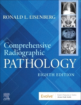
Comprehensive Radiographic Pathology
Seiten
2024
|
8th edition
Churchill Livingstone (Verlag)
978-0-443-12114-2 (ISBN)
Churchill Livingstone (Verlag)
978-0-443-12114-2 (ISBN)
Gain the pathology knowledge you need to produce quality radiographic images! A fully illustrated, easy-to-follow guide, Comprehensive Radiographic Pathology, 8th Edition describes the pathologies that can be diagnosed with medical imaging. The book provides an understanding of the principles of pathology and explains how to recognize the radiographic appearance of specific diseases and conditions. By mastering the suggested imaging techniques, you will produce the high-resolution images that lead to accurate diagnoses. Written by Ronald L. Eisenberg, an experienced radiologist and educator, this book will help you become a more competent professional and a valued contributor to the diagnostic team.
Radiographer Notes provide helpful suggestions for producing optimal radiographs of each organ system, and include information on positioning and exposure factor adjustments for patients with specific conditions or handling requirements.
Hundreds of high-quality illustrations cover all modalities, clearly demonstrating the clinical manifestations of different disease processes and providing a standard for the high-quality images needed in radiography practice.
Discussion of widely used imaging modalities includes the advantages and limitations of ultrasound, computed tomography (CT), magnetic resonance imaging (MRI), nuclear medicine, single-photon emission computed tomography (SPECT), positron emission tomography (PET), and fusion imaging.
Summary tables list the location and radiographic appearance of each disease as well as its treatment.
Recommended imaging procedures are provided for each pathologic condition, along with the sequence for choosing and performing alternative imaging studies if necessary.
Review questions help you assess your comprehension of the material in each chapter, with an answer key at the back of the book.
Thorough explanations provide an understanding of the pathologies that can be diagnosed with medical imaging.
Systems-based approach makes it easy to locate information and study one area at a time, helping you build an understanding of pathology.
NEW! Updated Radiographer Notes are included at the beginning of each chapter.
NEW! 30 new images are added, including CT scans pertaining to guidelines on detection of lung cancer.
NEW! Coverage of A.I. (artificial intelligence) and personalized medicine is added to this edition.
NEW! Coverage of COVID as pertaining to chest X-rays is added to this edition.
1 Introduction to Pathology
2 Specialized Imaging Techniques
3 Respiratory System
4 Skeletal System
5 Gastrointestinal System
6 Urinary System
7 Cardiovascular System
8 Nervous System
9 Hematopoietic System
10 Endocrine System
11 Reproductive System
12 Miscellaneous Diseases
| Erscheinungsdatum | 29.05.2024 |
|---|---|
| Verlagsort | London |
| Sprache | englisch |
| Maße | 216 x 276 mm |
| Gewicht | 1300 g |
| Themenwelt | Medizin / Pharmazie ► Gesundheitsfachberufe ► MTA - Radiologie |
| Medizinische Fachgebiete ► Radiologie / Bildgebende Verfahren ► Sonographie / Echokardiographie | |
| ISBN-10 | 0-443-12114-1 / 0443121141 |
| ISBN-13 | 978-0-443-12114-2 / 9780443121142 |
| Zustand | Neuware |
| Haben Sie eine Frage zum Produkt? |
Mehr entdecken
aus dem Bereich
aus dem Bereich
Begleitbuch für Sonografiekurse, Klinik und Praxis
Buch | Softcover (2023)
Urban & Fischer in Elsevier (Verlag)
27,00 €
Organbezogene Darstellung von Grund- und Aufbaukurs sowie …
Buch | Hardcover (2020)
Deutscher Ärzteverlag
99,99 €


