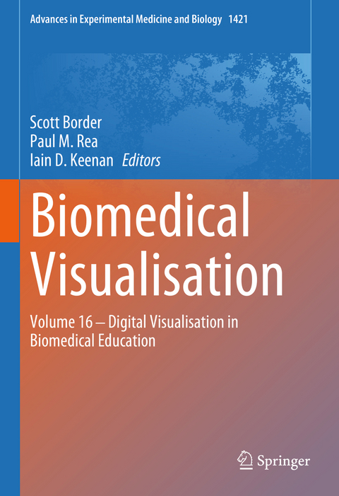
Biomedical Visualisation
Springer International Publishing (Verlag)
978-3-031-30378-4 (ISBN)
When studying medicine, healthcare, and medical sciences disciplines, learners are frequently required to visualise and understand complex three-dimensional concepts. Consequently, it is important that appropriate modalities are used to support their learning. Recently, educators have turned to new and existing digital visualisation approaches when adapting to pandemic-era challenges and when delivering blended post-pandemic teaching.
This book focuses on a range of key themes in anatomical and clinically oriented education that can be enhanced through visual understanding of the spatial three-dimensional arrangement and structure of human patients.The opening chapters describe important digital adaptations for the dissemination of biomedical education to the public and to learners. These topics are followed by reviews and reports of specific modern visualisation technologies for supporting anatomical, biomedical sciences, and clinical education. Examples include 3D printing, 3D digital models, virtual histology, extended reality, and digital simulation.
This book will be of interest to academics, educators, and communities aiming to modernise and innovate their teaching. Additionally, this book will appeal to clinical teachers and allied healthcare professionals who are responsible for the training and development of colleagues, and those wishing to communicate effectively to a range of audiences using multimodal digital approaches.
lt;p>Professor Scott Border B.Sc (Hons) N.T.F, F.A.S, S.F.H.E.A is Head of Anatomy, within The College of Medical, Veterinary and Life Sciences at the University of Glasgow. Professor Border trained as a behavioural neuroscientist, with an interest in the neural correlates of learning and memory. Having taught gross anatomy to medical students for 16 years, (mainly focussing on clinical neuroanatomy and head and neck anatomy) at Southampton, he is now leading anatomy education at the University of Glasgow. His main areas of interest focus on innovative approaches to anatomy education. Working with students as partners in anatomical education is a major investment of his time and this work has been recognised with a National Teaching Fellowship from Advance H.E in 2019.
Professor Paul M. Rea is Professor of Digital and Anatomical Education at the University of Glasgow. He is Director of Innovation, Engagement and Enterprise within the School of Medicine, Dentistry and Nursing. He is also a Senate Assessor for Student Conduct, Council Member on Senate and coordinates the day-to-day running of the Body Donor Program and is a Licensed Teacher of Anatomy, licensed by the Scottish Government.
He is qualified with a medical degree (MBChB), a MSc (by research) in craniofacial anatomy/surgery, a PhD in neuroscience, the Diploma in Forensic Medical Science (DipFMS), and an MEd with Merit (Learning and Teaching in Higher Education). He is a Senior Fellow of the Higher Education Academy, professional member of the Institute of Medical Illustrators (appointed recently as a Fellow of the Institute of Medical Illustrators) and a registered medical illustrator with the Academy for Healthcare Science.
Paul has published widely and presented at many national and international meetings, including invited talks. He has been the lead Editor for Biomedical Visualisation over 12 published volumes and is the founding editor for this book series. This has resulted in almost almost 90,000 downloads across these volumes, with contributions from over 400 different authors, across approximately 100 institutions from 19 countries across the globe. He is Associate Editor for the European Journal of Anatomy and has reviewed for 25 different journals/publishers. He is the Public Engagement and Outreach lead for anatomy coordinating collaborative projects with the Glasgow Science Centre, NHS and Royal College of Physicians and Surgeons of Glasgow. Paul is also a STEM ambassador and has visited numerous schools to undertake outreach work.
His research involves a long-standing strategic partnership with the School of Simulation and Visualisation The Glasgow School of Art. This has led to multi-million pound investment in creating world leading 3D digital datasets to be used in undergraduate and postgraduate teaching to enhance learning and assessment. This successful collaboration resulted in the creation of the world's first taught MSc Medical Visualisation and Human Anatomy combining anatomy and digital technologies. The Institute of Medical Illustrators also accredits it. It has created college-wide, industry, multi-institutional and NHS research linked projects for students.
Dr. Iain D. Keenan, B.Sc., (Hons.), Ph.D., M.Med.Ed., S.F.H.E.A., N.T.F., F.A.S is a Senior Lecturer in Anatomy within the School of Medicine, Newcastle University , U.K.. Iain has been interested in the visualisation of three-dimensional morphology since his PhD training and post-doctoral fellowships in developmental biology. Iain has since taught gross anatomy, histology, and embryology for medical, dental, physician associates, medical sciences and healthcare degree programmes at Newcastle University for more than 10 years. Iain has numerous leadership and mentoring roles in educational research and scholarship at Newcastle University and leads a programme of pedagogi
Part I. Communicating Visualisation.- Chapter 1. Science Communication and Biomedical Visualization - Two Sides of the Same Coin.- Chapter 2. Putting the Cart Before the Horse? Developing a Blended Anatomy Curriculum Supplemented by Cadaveric Anatomy.- Part II. Innovating Visualisation.- Chapter 3. The Third Dimension: 3D Printed Replicas and Other Alternatives to Cadaver-Based Learning.- Chapter 4. Evaluating a Photogrammetry-based Video for Undergraduate Anatomy Education.- Chapter 5. Virtual Microscopy Goes Global: The Images Are Virtual and the Problems Are Real.- Chapter 6. Online, Interactive, Digital Visualisation Resources That Enhance Histology Education.- Chapter 7. Leading Transformation in Medical Education Through Extended Reality.- Chapter 8. Visualisation Approaches in Technology-Enhanced Medical Simulation Learning: Current Evidence and Future Directions.- Chapter 9. Visualisation through Participatory/Interactive Theatre for the Health Sciences.
| Erscheinungsdatum | 02.08.2023 |
|---|---|
| Reihe/Serie | Advances in Experimental Medicine and Biology |
| Zusatzinfo | X, 203 p. 64 illus., 61 illus. in color. |
| Verlagsort | Cham |
| Sprache | englisch |
| Maße | 178 x 254 mm |
| Gewicht | 595 g |
| Themenwelt | Studium ► 1. Studienabschnitt (Vorklinik) ► Anatomie / Neuroanatomie |
| Schlagworte | 3D Printing • 3D visualization • anatomy • Anatomy Curriculum • augmented reality • clinical simulation • healthcare education • medical education • Medical Visualization Techniques • Teaching • Teaching Mental Health Nursing • Technology-Enhanced Learning • Videos • Virtual Histology |
| ISBN-10 | 3-031-30378-4 / 3031303784 |
| ISBN-13 | 978-3-031-30378-4 / 9783031303784 |
| Zustand | Neuware |
| Haben Sie eine Frage zum Produkt? |
aus dem Bereich


