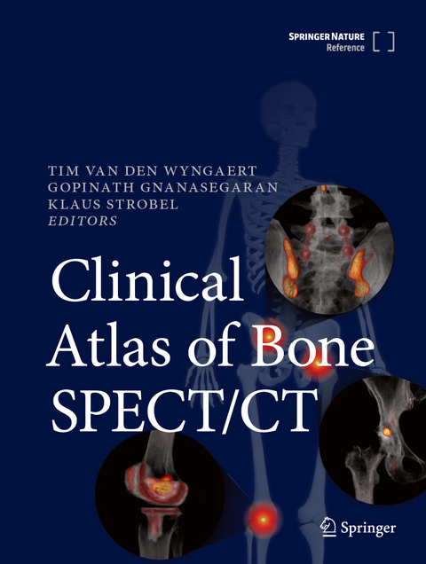
Clinical Atlas of Bone SPECT/CT
Springer International Publishing
978-3-031-26448-1 (ISBN)
It is published as part of the SpringerReference program, which delivers access to living editions constantly updated through a dynamic peer-review publishing process.
Tim Van den Wyngaert is a Professor at the University of Antwerp and a Senior Consultant in Nuclear Medicine at the Antwerp University Hospital (UZA), Belgium. He studied medicine and achieved his PhD at the University of Antwerp while also receiving clinical training in internal medicine and nuclear medicine at the Antwerp University Hospital (UZA). Professor Van den Wyngaert became a staff member and postdoctoral researcher at UZA in 2010, and a tenured faculty member in 2015. Professor Van den Wyngaert’s areas of interest include molecular imaging, bone disease, onco-osteology, and applied biomedical statistics. His current research interests include the clinical development of bone SPECT/CT in benign and malignant diseases, and the (pre)clinical validation of PET tracers for imaging the hallmarks of cancer. He is the Chairman of the European Association of Nuclear Medicine’s Bone and Joint Committee. Gopinath Gnanasegaran is a Consultant Nuclear Medicine Physician at the Royal Free London NHS Foundation Trust in London. Completed Nuclear Medicine residency at Guy’s Hospital (2007). He was appointed as a Consultant in Nuclear Medicine at Guys’ and St Thomas’ NHS Foundation Trust in 2007. He moved to Royal Free London NHS Foundation Trust in 2016. He was the Chair of Education Committee, British Nuclear Medicine Society (2013-2015) and Chair of Nuclear Medicine Special Interest Group, British Institute of Radiology (2012-2014). He was also a member of European Association of Nuclear Medicine, Bone & Joint Committee, and later member of EANM Oncology and Theranostics Committee. He is currently, Secretary General of World Federation of Nuclear Medicine & Biology. Dr Gnanasegaran has authored numerous papers in international, peer-reviewed journals. Klaus Strobel is adjunct Professor at University of Zurich, Switzerland and chief of Nuclear Medicine service at the department of Nuclear Medicine and Radiology at the Cantonal Hospital of Lucerne. He is doubly board certified in Radiology (2001) and Nuclear Medicine (2007) and trained amongst others at Nuclear Medicine department of University Hospital Zurich and MSK Radiology at University Hospital Balgrist. Prof. Strobel was vice-president of the European Association of Nuclear Medicine`s Bone and Joint Committee. His main research focus is bone and joint SPECT/CT and oncologic PET/CT. He served as teacher at International Diagnostic Course Davos (IDKD) and European School of Molecular Imaging an Therapy (ESMIT)
Sections:
1. SPECT/CT physics
2. SPECT/CT technical artefacts and pitfalls
3. Spine
4. Shoulder and Elbow
5. Hand and Wrist
6. Pelvis and Hip
7. Knee
8. Foot and Ankle
9. Sports injuries
10. Oncology
Chapters per section:
1. Normal X-ray and CT anatomy
2. Introduction to conditions and procedures
3. Bone SPECT/CT acquisition protocol
4. Pre-operative conditions
5. Post-operative conditions
| Erscheint lt. Verlag | 25.2.2024 |
|---|---|
| Zusatzinfo | XXIII, 1203 p. 942 illus., 612 illus. in color. In 2 volumes, not available separately. |
| Verlagsort | Cham |
| Sprache | englisch |
| Maße | 210 x 279 mm |
| Themenwelt | Medizinische Fachgebiete ► Chirurgie ► Unfallchirurgie / Orthopädie |
| Medizinische Fachgebiete ► Radiologie / Bildgebende Verfahren ► Nuklearmedizin | |
| Schlagworte | Anatomic correlations • Bone Disease • bone scintigraphy • Clinical Atlas of Bone SPECT/CT • Hybrid Bone Imaging • joint disease • Nuclear Medicine • Oncology • orthopaedics • Paediatrics • Post-operative assessment • Sports injuries |
| ISBN-10 | 3-031-26448-7 / 3031264487 |
| ISBN-13 | 978-3-031-26448-1 / 9783031264481 |
| Zustand | Neuware |
| Haben Sie eine Frage zum Produkt? |
aus dem Bereich

