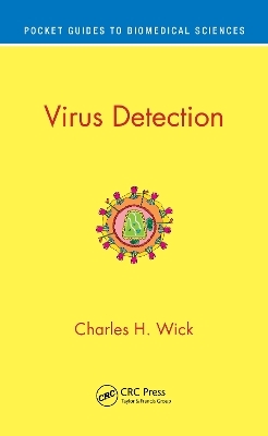
Virus Detection
CRC Press (Verlag)
978-0-367-61798-1 (ISBN)
Key Features
Covers common detection methods
Reviews the history of detection from antiquity to the present
Documents the strengths and weaknesses of various detection methods
Describes how to detect newly discovered viruses
Recommends specific applications for clinical, hospital, environmental, and public health uses
Charles H. Wick is a retired lieutenant colonel and senior scientist from the U.S. Army Edgewood Chemical Biological Center. He earned his PhD from the University of Washington. Dr. Wick is best known for work in forensic science done in concert with the Department of Defense. Dr. Wick’s research work has resulted in international recognition as an authority on individual performance for operations conducted on a nuclear, biological, and chemical battlefield. He has made lasting and important contributions to forensic science and antiterrorism and holds several U.S. patents in the area of microbe detection and classification. He has written more than 45 civilian and military publications and has received various awards and citations.
Abbreviations and Glossary 7
Preface 8
About the Author 11
Abstract 14
Virus Detection 15
Chapter 1 – Civilization and Disease 15
Chapter 2 – Microbes, Fungi, Bacteria, and Viruses 44
2.1 Fungi 47
2.1.1 What is a Fungus 47
2.1.2 How are Fungi detected/classified 47
2.2 Bacteria 48
2.2.1. What are Bacteria? 48
2.2.2 How are Bacteria detected and classified 48
2.3 Viruses 49
2.3.1 What are viruses? 49
2.3.2 How are Viruses detected and classified 50
Chapter 3 - Indirect Methods of Detecting viruses 63
Chapter 4 - Electron microscopy. 65
4.1 Transmission ission Electron microscopy 67
4.1.1 How does a Tranmission Electron Microscope (TEM) work? 67
4.1.2 How do you use Electron Microscopy 68
4.1.3 How do you identify a new virus 69
4.2 Scanning Electron microscopy 74
4.3 Examples of both TEM and SEM visualization of viruses. 77
Chapter 5 - Molecular Methods for detecting viruses 96
5.1 Polymerase Chain Reaction (PCR) 98
5.2 Antibody Methods 113
5.3 How do you add new viruses to the Antibody Method of detection? 115
Chapter 6 - Direct Virus Counting methods, such as IVDS. 116
6.1 How does direct counting work? 117
6.2 How do you identify a new virus with direct counting? 118
6.3 Why IVDS was invented 118
6.4 Flow chart showing how to use IVDS for virus detection 120
6.5 The Recommended uses of IVDS 124
6.6 Improving sensitivity of IVDS (Concentration and Accumulation) 129
6.7 An Example - Following COV-19 through 5 days and then a 3 Month follow-up 133
6.8 PCR and IVDS Compared 147
6.9 Summary of the Fielded IVDS 149
Chapter 7 – Mass Spectrometry Proteomic (MSP) Method 150
7.1 Introduction 151
7.2 Ion Mobility and Various Types of Mass Analyzers 154
7.2.1 Using Electrospray Ionization (ESI) method in detecting viruses 155
7.3 How do MSP Methods work for biological detection? 157
7.4 Detection and Identification of Viruses using MSP 159
7.5 Examples of Viruses Detected by MSP Methods using ABOid 162
7.5.1 African Swine Fever Virus (ASFV) - Variola Porcina 163
7.5.2 Alcelaphine Herpesvirus 1 (AlHV-1) 165
7.5.3 CamelpoxVirus (CMLV) 165
7.5.4 Cercopithecine herpesvirus 5 (CeHV-5) 166
7.5.5 Goatpox Virus Pellor (GTPV) 166
7.5.6 Lumpy Skin Disease Virus (LSDV) 166
7.5.7 MonkeypoxVirus Zaire-96-I-16 (MPV) 167
7.5.8 Sheeppox Virus 168
7.5.9 Vaccinia Virus (VACV) 168
7.5.10 Variola Virus (VARV) 168
7.5.11 Discussion of Viruses Detected by MSP and ABOid 169
7.6 Adding new viruses using MSP 170
7.7 Coronavirus Detection including SARS. 175
7.7.1 Coronavirus Viruses 176
7.7.2 National Average for Coronavirus 177
7.7.8 Verifying COVID-19 detection. 178
7.7.9 COVID-19 Detection Discussion 180
7.8 Summary of MSP-ABOid detection of viruses 180
Chapter 8 - Discussion 182
8.1 Introduction 184
8.2 What are the challenges when detecting viruses? 191
8.3 Clinical 194
8.4 Environmental 197
8.5 Using during a Pandemic 201
8.6 Other uses 203
References 208
| Erscheinungsdatum | 17.04.2023 |
|---|---|
| Reihe/Serie | Pocket Guides to Biomedical Sciences |
| Zusatzinfo | 12 Line drawings, color; 38 Line drawings, black and white; 15 Halftones, black and white; 12 Illustrations, color; 53 Illustrations, black and white |
| Verlagsort | London |
| Sprache | englisch |
| Maße | 133 x 203 mm |
| Gewicht | 200 g |
| Themenwelt | Medizin / Pharmazie ► Medizinische Fachgebiete ► Mikrobiologie / Infektologie / Reisemedizin |
| Studium ► Querschnittsbereiche ► Epidemiologie / Med. Biometrie | |
| Naturwissenschaften ► Biologie ► Genetik / Molekularbiologie | |
| ISBN-10 | 0-367-61798-6 / 0367617986 |
| ISBN-13 | 978-0-367-61798-1 / 9780367617981 |
| Zustand | Neuware |
| Informationen gemäß Produktsicherheitsverordnung (GPSR) | |
| Haben Sie eine Frage zum Produkt? |
aus dem Bereich


