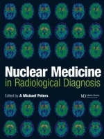
Nuclear Medicine In Radiological Diagnosis
Taylor & Francis Ltd (Verlag)
978-1-899066-50-6 (ISBN)
- Titel ist leider vergriffen;
keine Neuauflage - Artikel merken
A Michael Peters MA MD MSc FRCPath FRCP FRCR FMedSci, Professor of Nuclear Medicine and Honorary Consultant, Department of Nuclear Medicine, Addenbrooke's Hospital, Cambridge, UK
Section I: Muscoloskeletal System. Back Pain. Imaging of Malignant Disorders of Bone. The Painful Prosthetic Joint. Infection in the Peripheral Skeleton. Unexplained Pain and Trauma in the Peripheral Skeleton. Metabolic Bone Disease. Painful and/or Swollen Joint. Pediatric Bone Scanning. Skeletal Trauma. Section II: Nephrourology. Measuring Renal Function. Urinary Dilation and Obstruction in the Adult. Imaging Children Born with an Abnormal Urinary Tract. Urinary Tract Infection in Children. Clinicians' Requirements of Radiology when Investigating Patients with Hypertension. Hypertension and Suspected Renovascular Disease. Assessment in Renal Transplantation. Imaging of Acute Renal Failure. Section III: Gastrointestinal System. Evaluation of the Liver and Biliary Tree. Radionucleide Imaging in Gastrointestinal Bleeding. Radiological Investigation of Inflammatory Bowel Disease. Gastrointestinal Tract. Section IV: Cardiology. What the Cardiologist Requires of Nuclear Medicine. Evaluation of the Patient with Chest Pain: Rationale for the Use of Radionuclide Imaging. Imaging the Heart After Myocardial Infarction. Imaging the Heart in Patients with Cardiac Failure. Imaging Techniques in Determining Myocardial Viability and Hibernation. Clinical Application of Radionucleide Imaging in Cardiomyopathies and Valvular Diseases. Clinical Value of Measuring Right Ventricular Function. Section V: Neurology. Nuclear Medicine Techniques in the Investigation of Dementia. Functional Imaging in Brain Tumors. Nuclear Medicine in the Psychoses. Functional Imaging of Movement Disorders. Epilepsy and Related Seizure Disorders. The Role of SPECT in Stroke. Section VI: Hematology. Nuclear Medicine in Myeloproliferative Disease. Radioisotope Investigation of Chronic Anemia. Investigating Thrombocytopenia. The Spleen. Positron Emission Tomography in Hematology. Lymphoproliferative Disorders. The Swollen Limb. Section VII: The Lung. Imaging the Legs in Patients with Suspected Pulmonary Embolism. Ventilation-Perfusion Imaging for a Venous Thrombo-embolism: A Changing Role in the Diagnostic Algorithm. Clinical Role of Ventilation-perfusion Scanning in Non-embolic Lung Disease. Role of Gallium Scanning in the Management of Chronic Lung Disease. PET Imaging in Lung Cancer. Clinical Applications of DTPA Aerosol Clearance. Lung Scanning in Pediatrics. Section VIII: Endocrinology. Approach to the Patient with a Thyroid Nodule. Nuclear Medicine in the Management of the Patient with Abnormal Thyroid Function. Management of Goiter. Imaging the Parathyroids. Imaging the Adrenal Cortex. Section IX: Infection/Inflammation. Undiagnosed Fever. AIDS, Infection and Nuclear Medicine. Imaging Intra-abdominal Sepsis. Section X: Oncology. Radioimmunoscintigraphy. Finding Pheochromocytoma. Clinical Applications of PET in Oncology. Imaging the Breast. Management of Thyroid Carcinoma.
| Erscheint lt. Verlag | 5.6.2003 |
|---|---|
| Zusatzinfo | 80 Illustrations, color; 720 Illustrations, black and white |
| Verlagsort | London |
| Sprache | englisch |
| Maße | 216 x 279 mm |
| Gewicht | 2948 g |
| Themenwelt | Medizinische Fachgebiete ► Radiologie / Bildgebende Verfahren ► Nuklearmedizin |
| Medizinische Fachgebiete ► Radiologie / Bildgebende Verfahren ► Radiologie | |
| ISBN-10 | 1-899066-50-0 / 1899066500 |
| ISBN-13 | 978-1-899066-50-6 / 9781899066506 |
| Zustand | Neuware |
| Haben Sie eine Frage zum Produkt? |
aus dem Bereich


