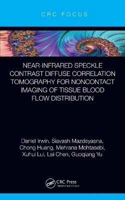
Near-infrared Speckle Contrast Diffuse Correlation Tomography for Noncontact Imaging of Tissue Blood Flow Distribution
CRC Press (Verlag)
978-1-032-13387-4 (ISBN)
Imaging of tissue blood flow (BF) distributions provides vital information for the diagnosis and therapeutic monitoring of various vascular diseases. The innovative near-infrared speckle contrast diffuse correlation tomography (scDCT) technique produces full 3D BF distributions. Many advanced features are provided over competing technologies including high sampling density, fast data acquisition, noninvasiveness, noncontact, affordability, portability, and translatability across varied subject sizes. The basic principle, instrumentation, and data analysis algorithms are presented in detail. The extensive applications are summarized such as imaging of cerebral BF (CBF) in mice, rat, and piglet animals with skull penetration into deep brain. Clinical human testing results are described by recovery of BF distributions on preterm infants (CBF) through incubator wall, and on sensitive burn tissues and mastectomy skin flaps without direct device-tissue interactions. Supporting activities outlined include integrated capability for acquiring surface curvature information, rapid 2D blood flow mapping, and optimizations via tissue-like phantoms and computer simulations. These applications and activities both highlight and guide the reader as to the expected abilities and limitations of scDCT for adapting into their own preclinical/clinical research, use in constrained environments (i.e., neonatal intensive care unit bedside), and use on vulnerable subjects and measurement sites.
Lei Chen is an Associate Professor in the Department of Physiology, Spinal Cord and Brain Injury Research Center, and the Department of Biomedical Engineering at the University of Kentucky. His research focuses on the activation of stem cells and other reparative mechanisms after a head injury, including ischemic stroke, head trauma and neonatal intraventricular hemorrhage. Those animal models have been utilized for the validation of scDCT to achieve the continuous and longitudinal monitoring of cerebral injury and recovery. Chong Huang worked as a Postdoctoral Scholar and then Research Assistant Professor in the Department of Biomedical Engineering at the University of Kentucky. Over the past nine years, he worked on the development of various near-infrared diffuse optical spectroscopic and tomographic technologies for noninvasive assessment of tissue hemodynamics and metabolism. He is the co-inventor of the scDCT technology. Daniel Irwin is a Research Scientist in the Department of Biomedical Engineering at the University of Kentucky. He has been contributing to the Biomedical Optics field for nine years, completing his PhD in 2018 with dissertation work including efforts towards scDCT development, analysis, and imaging. His current research involves advancing near-infrared diffuse speckle-based spectroscopic and imaging instrumentation. Xuhui Liu is a PhD candidate in the Department of Biomedical Engineering at the University of Kentucky. He obtained his bachelor’s degree in Electrical Engineering at the University of Electronic Science and Technology of China. His research focuses on developing a wearable fiber-free optical sensor to continuously monitor cerebral blood flow and oxygenation in different subjects (mice, piglets, preterm infants). Siavash Mazdeyasna received his PhD in Biomedical Engineering from the University of Kentucky (UK) in the fall of 2020. His research during his doctoral and postdoctoral studies at UK has provided him with extensive experience in biomedical engineering especially in developing the state-of-the-art diffuse optical imaging system. He has worked on both hardware and software development, data analysis, and validations in tissue-simulating phantoms, small animals, and human subjects. Mehrana Mohtasebi is a PhD candidate in the Department of Biomedical Engineering at the University of Kentucky. Over the past three years, Mehrana has been working on optimization and validation of scDCT against established techniques for quantifying resting-state functional connectivity networks in animal models of stroke and intraventricular hemorrhage to provide objective assessments of brain tissue injury. Guoqiang Yu is a Professor in the Department of Biomedical Engineering at the University of Kentucky. Over the past decade, he leads the Biomedical Optics Laboratory where his group has invented several highly powerful functional imaging technologies, including scDCT for noncontact imaging of brains, burns, wounds, and reconstructive tissues, a wearable fiber-free optical sensor for continuous cerebral monitoring in freely behaving subjects, wearable fluorescence eye loupes, and a time-resolved laser speckle contrast imaging system. These patented technologies and devices hold great promise for imaging numerous organs in preclinical and clinical settings. He has established a track record of publications in high impact journals. Particularly, he has written a book chapter in the Handbook of Biomedical Optics (2011, CRC Press), titled "Near-infrared diffuse correlation spectroscopy (DCS) for assessment of tissue blood flow".
1. Introduction 2. scDCT Methods 3. scDCT Validations and Applications 4. Summary and future perspectives
| Erscheinungsdatum | 06.08.2022 |
|---|---|
| Zusatzinfo | 1 Tables, black and white; 3 Line drawings, black and white; 17 Halftones, black and white; 20 Illustrations, black and white |
| Verlagsort | London |
| Sprache | englisch |
| Maße | 138 x 216 mm |
| Gewicht | 460 g |
| Themenwelt | Medizinische Fachgebiete ► Radiologie / Bildgebende Verfahren ► Computertomographie |
| Technik ► Umwelttechnik / Biotechnologie | |
| ISBN-10 | 1-032-13387-2 / 1032133872 |
| ISBN-13 | 978-1-032-13387-4 / 9781032133874 |
| Zustand | Neuware |
| Informationen gemäß Produktsicherheitsverordnung (GPSR) | |
| Haben Sie eine Frage zum Produkt? |
aus dem Bereich


