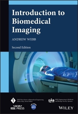
Introduction to Biomedical Imaging
Wiley-IEEE Press (Verlag)
978-1-119-86771-5 (ISBN)
In the newly revised second edition of Introduction to Biomedical Imaging, distinguished researcher Dr. Andrew Webb delivers a comprehensive description of the fundamentals and applications of the most important current medical imaging techniques: X-ray and computed tomography, nuclear medicine, ultrasound, magnetic resonance imaging, and various optical-based methods. Each chapter explains the physical principles, instrument design, data acquisition, image reconstruction, and clinical applications of its respective modality.
This latest edition incorporates descriptions of recent developments in photon counting CT, total body PET, superresolution-based ultrasound, phased-array MRI technology, optical coherence tomography, and iterative and model-based image reconstruction techniques. The final chapter discusses the increasing role of artificial intelligence/deep learning in biomedical imaging. The text also includes a thorough introduction to general image characteristics, including discussions of signal-to-noise and contrast-to-noise.
Perfect for graduate and senior undergraduate students of biomedical engineering, Introduction to Biomedical Imaging, 2nd Edition will also earn a place in the libraries of medical imaging professionals with an interest in medical imaging techniques.
ANDREW WEBB, PHD, is a full Professor and Director of the C.J. Gorter Centre for High Field Imaging in the Department of Radiology at Leiden University Medical Center in the Netherlands. He is also a faculty member in Electrical Engineering at TU Delft and holds an adjunct position at the Beckman Institute for Advanced Science and Technology at the University of Illinois. He published the first edition of this title with Wiley in 2003.
Preface xv
Introduction xix
About the Companion Website xxxi
1 Image and Imaging System Characteristics 1
1.1 General Image and Imaging System Characteristics 1
1.2 Concept of Spatial Frequency 2
1.3 Spatial Resolution 3
1.3.1 Imaging System Point Spread Function 4
1.3.2 Imaging System Resolving Power 5
1.3.3 Imaging System Modulation Transfer Function 6
1.4 Signal-to-Noise Ratio 7
1.5 Contrast-to-Noise Ratio 9
1.6 Signal Digitization: Dynamic Range and Resolution 9
1.7 Post-acquisition Image Filtering 11
1.8 Assessing the Clinical Impact of Improvements in System Performance 12
1.8.1 The Receiver Operating Characteristic Curve 13
1.A.1 Fourier Transforms 14
1.A.2 Fourier Transforms of Time Domain and Spatial Frequency Domain Signals 15
1.A.3 Useful Properties of the Fourier Transform 16
Exercises 17
References 19
Further Reading 20
2 X-ray Imaging and Computed Tomography 23
2.1 General Principles of Imaging with X-rays 23
2.2 X-ray Production 25
2.2.1 The X-ray Tube 25
2.2.2 The X-ray Energy Spectrum 29
2.3 Interactions of X-rays with Tissue 32
2.3.1 Compton Scattering 33
2.3.2 The Photoelectric Effect 34
2.4 Linear and Mass Attenuation Coefficients of X-rays in Tissue 36
2.5 Instrumentation for Planar X-ray Imaging 38
2.5.1 Collimator 38
2.5.2 Anti-scatter Grid 38
2.6 Digital X-ray Detectors 40
2.7 X-ray Image Characteristics 42
2.7.1 Signal-to-Noise 42
2.7.2 Spatial Resolution 44
2.7.3 Contrast-to-Noise 45
2.8 X-ray Contrast Agents 46
2.8.1 Contrast Agents for the Gastrointestinal Tract 46
2.8.2 Iodine-Based Contrast Agents 46
2.9 X-ray Imaging Methods 47
2.9.1 X-ray Fluoroscopy 48
2.9.2 Digital Subtraction Angiography 48
2.10 Clinical Applications of X-ray Imaging 49
2.10.1 Digital Mammography 49
2.10.2 Abdominal X-ray Scans 50
2.11 Computed Tomography 51
2.12 CT Scanner Instrumentation 53
2.12.1 Beam Filtration 55
2.12.2 Detectors for Computed Tomography 56
2.13 Image Processing for Computed Tomography 57
2.13.1 Filtered Backprojection (FBP) Techniques 57
2.13.2 Fan-Beam and Spiral Reconstructions 61
2.14 Iterative Algorithms 63
2.15 Radiation Dose 65
2.16 Spectral/Dual Energy CT 66
2.17 Photon-Counting CT 69
2.18 Cone Beam, Mobile, and Portable CT Units 71
2.19 Clinical Applications of Computed Tomography 72
2.19.1 Head and Neurovascular Scans 72
2.19.2 Pulmonary Disease 73
2.19.3 Abdominal Imaging 73
2.19.4 Cardiovascular Imaging 74
Exercises 75
References 81
Further Reading 82
3 Nuclear Medicine 85
3.1 General Principles of Nuclear Medicine 85
3.2 Radioactivity and Radiotracer Half-life 87
3.3 Common Radiotracers Used for SPECT 89
3.4 The Technetium Generator 90
3.5 The Distribution of Technetium-Based Radiotracers within the Body 92
3.6 Instrumentation for SPECT and SPECT/CT 94
3.6.1 Collimators 94
3.6.2 Scintillation Crystal and Photomultiplier Tube-Based Detectors 98
3.6.3 The Anger Position Network and Pulse Height Analyzer 100
3.6.4 Solid-State Detectors and Specialized Cardiac Scanners 102
3.7 Image Reconstruction 103
3.7.1 Attenuation Correction 104
3.7.2 Scatter Correction 105
3.8 Image Characteristics 106
3.8.1 Signal-to-Noise 106
3.8.2 Spatial Resolution 107
3.8.3 Contrast-to-Noise 107
3.9 Clinical Applications of SPECT 107
3.9.1 Brain Imaging 108
3.9.2 Bone Scanning and Tumor Detection 108
3.9.3 Cardiac Imaging 110
3.9.4 The Respiratory System 110
3.9.5 The Liver and Reticuloendothelial System 112
3.10 Positron Emission Tomography 113
3.11 Radiotracers Used for PET 115
3.12 Instrumentation for PET 116
3.12.1 Scintillation Crystals and Detector Electronics 117
3.13 Image Reconstruction 118
3.13.1 Annihilation Coincidence Detection and Removal of Accidental Coincidences 119
3.13.2 Attenuation Correction 120
3.13.3 Scatter Correction 120
3.13.4 Dead-Time Correction 120
3.14 Image Characteristics 121
3.14.1 Spatial Resolution 121
3.14.2 Signal-to-Noise 121
3.14.3 Contrast-to-Noise 122
3.15 Acquisition Methods for PET 122
3.16 Total Body PET Systems 122
3.17 Clinical Applications of PET/CT 124
3.17.1 Body Oncology 124
3.17.2 Brain Imaging 125
3.17.3 Cardiac Imaging 125
Exercises 126
References 131
Further Reading 132
4 Ultrasound Imaging 135
4.1 General Principles of Ultrasound Imaging 135
4.2 Wave Propagation and Acoustic Impedance 137
4.3 Wave Reflection 139
4.4 Energy Loss Mechanisms in Tissue 142
4.4.1 Scattering 142
4.4.2 Absorption 143
4.4.3 OverallWave Attenuation 145
4.5 Instrumentation 145
4.5.1 Transducer Construction 146
4.5.2 Transducer Arrays 149
4.5.2.1 Linear Sequential Array 151
4.5.2.2 Curvilinear/Convex Sequential Array 151
4.5.2.3 Linear-Phased Array 152
4.6 Signal Detection and Processing 153
4.6.1 Time Gain Compensation 153
4.6.2 Receive Beam Forming 154
4.7 Diagnostic Scanning Modes 155
4.7.1 A-Mode, M-Mode, and B-Mode Scans 155
4.7.2 Three-Dimensional Imaging 156
4.7.3 Compound Imaging 156
4.7.4 Other Transmit and Receive Beamforming Techniques 158
4.8 Image Characteristics 158
4.8.1 Signal-to-Noise 158
4.8.2 Spatial Resolution 159
4.8.2.1 Axial Resolution 159
4.8.2.2 Lateral Resolution 160
4.8.3 Contrast-to-Noise 161
4.9 Artifacts in Ultrasound Imaging 161
4.10 Blood Velocity Measurements Using Ultrasound 163
4.10.1 The Doppler Effect 163
4.10.2 Pulsed-Mode Doppler Measurements 164
4.10.3 Color Doppler/B-mode Duplex and Triplex Imaging 167
4.10.4 ContinuousWave Doppler (CWD) Measurements 168
4.11 Ultrasound Contrast Agents 169
4.11.1 Harmonic and Pulse Inversion Techniques 171
4.11.2 Super-Resolution in Ultrasound Imaging 172
4.12 Safety and Bioeffects in Ultrasound Imaging 174
4.13 Point-of-Care Ultrasound Systems 175
4.14 Clinical Applications of Ultrasound 176
4.14.1 Obstetrics and Gynecology 176
4.14.2 Breast Imaging 176
4.14.3 Musculoskeletal Structure 177
4.14.4 Abdominal 178
Exercises 179
References 185
Further Reading 186
5 Magnetic Resonance Imaging 189
5.1 General Principles of MRI Acquisition and Hardware 189
5.2 Nuclear Magnetization 191
5.2.1 Quantum Mechanical Description 191
5.2.2 Classical Description 195
5.2.3 Hydrogen Nuclei inWater and Lipid 197
5.2.4 Radiofrequency Pulses and the Creation of Transverse Magnetization 197
5.2.5 Signal Detection and Fourier Transformation 199
5.3 T1 and T2 Relaxation Mechanisms and Tissue Relaxation Times 200
5.3.1 Tissue-Dependent Relaxation Times 202
5.3.2 Measurement of T1 and T2: Inversion-Recovery and Spin-Echo Sequences 204
5.4 The MR Free Induction Decay 206
5.5 Magnetic Resonance Imaging 207
5.5.1 Spatial Localization 207
5.5.2 Imaging Concepts 209
5.5.2.1 Slice Selection 210
5.5.2.2 Phase-encoding 212
5.5.2.3 Frequency-encoding 214
5.5.2.4 The k-Space Formalism and Image Reconstruction 214
5.6 Imaging Sequences and Techniques 217
5.6.1 Multislice Gradient-Echo Sequences 217
5.6.2 Multislice Spin-Echo and Turbo-Spin-Echo Sequences 219
5.6.3 Three-Dimensional Gradient-Echo and Spin-Echo Sequences 221
5.6.4 Proton Density, T1-, T2-, and T∗2 -Weighted Sequences 222
5.6.5 Lipid Suppression Techniques 223
5.7 MRI Contrast Agents 226
5.8 Advanced Sequences 228
5.8.1 Magnetic Resonance Angiography 228
5.8.2 Diffusion-Weighted Imaging with Echo Planar Readout 230
5.8.3 In Vivo Localized Spectroscopy 232
5.8.4 Functional MRI 233
5.9 Instrumentation 235
5.9.1 Magnet Design 235
5.9.1.1 Clinical Superconducting Magnets 235
5.9.1.2 Very High Field Magnets 238
5.9.1.3 High-Temperature Superconductors 239
5.9.1.4 Mid- and Low-Field Magnets 239
5.9.2 Magnetic Field Gradient Coils 240
5.9.3 Radiofrequency Coils 244
5.9.3.1 Transmit Coil 244
5.9.4 Receiver Coil Array 244
5.9.5 Receiver Electronics 246
5.10 Image Reconstruction from Undersampled Data 247
5.10.1 Parallel Imaging Using an Array of Receiver Coils 248
5.10.2 Compressed Sensing 250
5.11 Image Characteristics 252
5.11.1 Signal-to-Noise 252
5.11.2 Spatial Resolution 254
5.11.3 Contrast-to-Noise 254
5.12 Image Artifacts 255
5.13 RF Safety Considerations 256
5.14 Clinical Applications of MRI 257
5.14.1 Neurological 258
5.14.2 Body Imaging 259
5.14.3 Musculoskeletal 259
5.14.4 Cardiac 259
Exercises 262
References 274
Further Reading 277
6 Optical Imaging 279
6.1 General Properties of Optical Imaging Methods 279
6.2 Propagation of Light Through Tissue 281
6.3 Body Emissivity Techniques – Infrared Thermography 284
6.4 Direct Imaging with Visible Light 285
6.4.1 Fundus Photography 285
6.4.2 Scheimpflug Camera 287
6.5 Optical Coherence Tomography (OCT) 288
6.5.1 Basic Principles of Interferometry 289
6.5.2 Instrumentation for OCT 291
6.5.2.1 Light Sources 291
6.5.2.2 Beam-Splitter 292
6.5.2.3 Photodetectors 292
6.5.3 Image Characteristics of OCT 292
6.5.4 OCT Angiography 294
6.5.5 Clinical Applications of OCT 295
6.6 Fluorescence-Guided Surgery (FGS) 296
6.6.1 Principle of Fluorescence 296
6.6.2 Fluorescent Probes 296
6.6.3 Instrumentation for Fluorescence Imaging 297
6.6.4 Clinical Applications of Fluorescence-Guided Surgery 298
6.7 Near-Infrared Spectroscopy (NIRS) and Diffuse Optical Tomography (DOT) 299
6.7.1 Principle of NIRS 299
6.7.2 Instrumentation for NIRS 301
6.7.3 Principle of DOT 301
6.7.4 Clinical Applications of DOT 302
6.8 Photoacoustic Imaging (PAI) 303
6.8.1 Principles of PAI 303
6.8.2 Photoacoustic Microscopy and Photoacoustic Computed Tomography 304
6.8.3 Instrumentation for PAI 305
6.8.4 Clinical Applications of PAI 305
References 306
Further Reading 308
7 Artificial Intelligence 311
7.1 Artificial Intelligence in Biomedical Imaging 311
7.2 Artificial Intelligence, Machine Learning, Deep Learning, and Neural Networks 312
7.2.1 Neural Networks 313
7.3 Deep Learning in Image Reconstruction 317
7.4 Convolutional Neural Networks (CNNs) 318
7.5 Artificial Intelligence in X-ray and CT 321
7.5.1 Image Reconstruction 321
7.5.2 Clinical Applications 322
7.6 Artificial Intelligence in SPECT and PET 323
7.6.1 Image Reconstruction 323
7.6.2 Clinical Applications 324
7.7 Artificial Intelligence in Ultrasound 324
7.7.1 Improved Data Acquisition 325
7.7.2 Image Post-processing 326
7.7.3 Image Analysis and Clinical Applications 326
7.8 Artificial Intelligence in MRI 326
7.8.1 Image Reconstruction 326
7.8.2 Clinical Applications 328
7.9 Artificial Intelligence in Optical Imaging 329
7.10 AI and Radiomics 329
7.11 Challenges for AI in Biomedical Imaging 330
References 331
Further Reading 337
Index 341
| Erscheinungsdatum | 06.10.2022 |
|---|---|
| Reihe/Serie | IEEE Press Series on Biomedical Engineering |
| Sprache | englisch |
| Gewicht | 776 g |
| Themenwelt | Informatik ► Theorie / Studium ► Künstliche Intelligenz / Robotik |
| Medizin / Pharmazie ► Medizinische Fachgebiete | |
| ISBN-10 | 1-119-86771-1 / 1119867711 |
| ISBN-13 | 978-1-119-86771-5 / 9781119867715 |
| Zustand | Neuware |
| Haben Sie eine Frage zum Produkt? |
aus dem Bereich


