Atlas of Dermoscopy
CRC Press (Verlag)
978-1-138-59598-9 (ISBN)
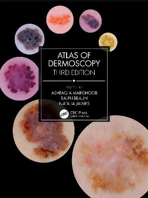
The much awaited third edition of the leading reference book in dermoscopy has undergone comprehensive revisions to all chapters, with updates and expanded content providing the reader with a more comprehensive and in-depth coverage of skin conditions, ranging from skin neoplasia to hair, nails, infections and inflammatory diseases. This compilation of contemporary dermoscopy knowledge will benefit the novice in building expertise and benefit experienced practitioners also by providing nuanced insights. Prepare to embark on a journey of learning as leading international experts in the field of dermoscopy explain via text and annotated exemplar images the amazing world of subsurface cutaneous colors and structures visible through a dermatoscope!
Ashfaq Marghoob MD is a Dermatologist at Memorial Sloan-Kettering Cancer Centers in Manhattan and is the Director of the outpatient Memorial Sloan-Kettering Skin Cancer Center in Hauppauge, New York, USA. Ralph Braun MD, PhD, is a Dermatologist at University Hospital Zürich, Switzerland. Natalia Jaimes MD is a dermatologist at the Dr Phillip Frost Department of Dermatology and Cutaneous Surgery, and Sylvester Comprehensive Cancer Center, University of Miami Miller School of Medicine, Miami, Florida, USA
Contributors. Foreword. Introduction. Principles of Dermoscopy and Dermoscopic Equipment. Histopathologic Correlations of Dermoscopic Structures. Primary Diagnostic Algorithms: Pattern Analysis Revised. Primary Diagnostic Algorithms: Top-Down 2-Step Pattern Analysis Algorithm. Primary Diagnostic Algorithms: A Decision Algorithm for Nonpigmented Skin Lesions. Triage Algorithms: A Decision Algorithm for Pigmented Lesions Based on Revised Pattern Analysis (“Chaos and Clues”). Triage Algorithms: Triage Amalgamated Dermoscopy Algorithm (TADA). Triage Algorithms: Other Dermoscopic Algorithms in Skin Cancer Triage. Nonmelanocytic lesions: Dermatofibroma. Nonmelanocytic Lesions: Basal Cell Carcinoma. Nonmelanocytic Lesions: Actinic Keratosis. Nonmelanocytic Lesions: Actinic Keratosis, Bowen’s Disease, Keratoacanthoma, and Squamous Cell Carcinoma. Nonmelanocytic Lesions: Solar Lentigines, Seborrheic Keratoses, and Lichen Planus–Like Keratosis. Nonmelanocytic Lesions: Vascular Lesions. Nonmelanocytic Lesions: Clear Cell Acanthoma, Poroma, Sebaceous Hyperplasia, and Other Adnexal Neoplasms. Benign Melanocytic Lesions: Congenital Melanocytic Nevi. Benign Melanocytic Lesions: Melanocytic Nevi. Benign Melanocytic Lesions: Intradermal Nevus. Benign Melanocytic Lesions: Blue Nevi and Variants. Benign Melanocytic Lesions: Spitz and Reed Nevi. Benign Melanocytic Lesions: Other Nevi. Melanocytic Lesions: Superficial Spreading Melanomas. Melanocytic Lesions: Nodular Melanoma. Melanocytic Lesions: Lentigo Maligna or Lentiginous Melanoma on Sun-Damaged Skin. Melanocytic Lesions: Acrolentiginous Melanoma. Melanocytic Lesions: Other Melanoma Subtypes. Melanocytic Lesions: Amelanotic and Hypomelanotic Melanoma. Methods to Differentiate Nevi from Melanoma: Pattern Analysis and Melanoma-Specific Criteria. Methods to Differentiate Nevi from Melanoma: Rules and Algorithms. Methods to Differentiate Nevi from Melanoma: Rules to Avoid Missing a Melanoma. Methods to Differentiate Nevi from Melanoma: Methods to Improve Sensitivity and Specificity in Melanoma Diagnosis. Collision Tumors and Exceptions to Rules: False Positive and False Negative. Special Locations: Face. Special Locations: Palms and Soles (Volar Surface). Special Locations: Mucosal Surfaces and Glabrous Skin on Glans and Labia. Special Locations: Nails. Special Locations: Hair and Scalp (Trichoscopy). Dermoscopy in General Dermatology: Infectious Diseases. Dermoscopy in General Dermatology: Inflammatory Dermatoses (Inflammoscopy). Digital Monitoring: Short- and Long-Term. Index.
| Erscheinungsdatum | 19.08.2022 |
|---|---|
| Zusatzinfo | 43 Tables, black and white; 68 Line drawings, color; 1357 Halftones, color; 1425 Illustrations, color |
| Verlagsort | London |
| Sprache | englisch |
| Maße | 210 x 280 mm |
| Gewicht | 1220 g |
| Themenwelt | Medizin / Pharmazie ► Allgemeines / Lexika |
| Medizin / Pharmazie ► Medizinische Fachgebiete ► Dermatologie | |
| Studium ► 2. Studienabschnitt (Klinik) ► Pathologie | |
| Studium ► Querschnittsbereiche ► Prävention / Gesundheitsförderung | |
| ISBN-10 | 1-138-59598-5 / 1138595985 |
| ISBN-13 | 978-1-138-59598-9 / 9781138595989 |
| Zustand | Neuware |
| Informationen gemäß Produktsicherheitsverordnung (GPSR) | |
| Haben Sie eine Frage zum Produkt? |
aus dem Bereich


