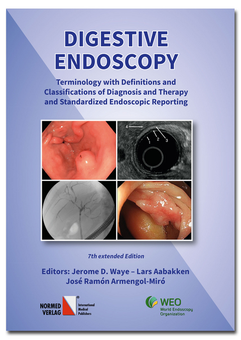
Digestive Endoscopy
Normed (Verlag)
978-3-89199-100-8 (ISBN)
Principles of endoscopic terminology
There are three steps in the formulation of an endoscopic statement:
1. description,
2. interpretation,
3. final diagnosis including the result of histological or cytological examination.
1. Description
As with every procedure, endoscopy must use appropriate terms to describe the findings.
Being essentially a visual method analogous to dermatology, endoscopy describes macroscopic
features of the inside of the digestive tube and abdominal cavity, especially the surface and the
colour of the mucosa or serosa, the movements of the wall, the shape and appearance of
lesions. The descriptive part must, therefore, use only terms related to such macroscopic
features. It is inappropriate to use terms which imply features which cannot be established
by naked-eye examination, e.g. histological structure such as “gastritis” or “colitis”, or can be
only partly or roughly evaluated e.g. depth of a lesion, chronicity etc.
Descriptive endoscopic terminology, therefore, is to a large extent specific for endoscopy and
diVerent from other methods and disciplines such as pathology.
As has been emphasized in the preface, the subject of this treatise is terminology of endoscopic
findings, not the terminology of digestive diseases.
2. Interpretation
The interpretation of the findings in the sense of a clinical diagnosis is expressed in the summary
of the report. It should explicitly contribute to answering the question raised by the
indication for examination. Whilst the description must be as objective as possible the interpretation
of the findings depends to a large extent on the views of the examining endoscopist.
3. Final diagnosis
In many cases a complementary method such as biopsy is used to establish the final diagnosis.
On this basis the WEO terminology does not seek to compete with diagnostic classification
of diseases such as the ICD Diagnostic Codes [17].
Selection of appropriate descriptive terms
Endoscopic terms must avoid, as far as possible, histopathological or clinical terms which
cannot be identified endoscopically with certainty. Some commonly used terms for lesions
and diseases which can be recognised endoscopically with reasonable accuracy can however
be used as an alternative terms., e.g. ulcer (for defect), hyperemia (for red mucosa). When
descriptive terms are not available, some new words are proposed in this book (e.g. “nodule”
instead of “chronic erosion”).
The term tumor in endoscopy is used to describe a protrusion considered most probably to
be of neoplastic nature; however, this term is not used in case of protrusions due to a distinct
entity such as a lipoma. The term mass is often used instead of tumor.
In place of inappropriate or ambiguous terms new words had be selected and the editors have
strived to make them as simple and self-descriptive as possible. Examples of the quest for
simplicity can be shown in the terms gastritis and erosion.
Gastritis is essentially an histologic finding whereas its endoscopic characteristics – in contrast
to esophagitis and colitis –are inconspicuous or unreliable. Accordingly, descriptive terms
(red, congested, erythematous etc. mucosa) are be used when noting changes in the gastric
mucosa. However, the term atrophic gastritis is an exception since gastric atrophy – an advanced
stage of chronic gastritis – can be recognized endoscopically with reasonable certainly.
The term gastropathy should be used to describe some endoscopically well characterized
non inflammatory conditions for which the term gastritis has often been used inappropriately
(Appendix 3B).
Erosion also is defined histologically so this term should be used with caution by endoscopists.
Most erosions are microscopic lesions and cannot be identified endoscopically unless some
special method is used (magnification, vital staining). In many diVerent conditions erosion is
an epiphenomenon which has little or nothing to do with the endoscopic aspect of the lesion
[18].
The ambiguity of the term erosion, as used in endoscopy, is due to the fact that it designates
lesions of diVerent appearance and character. Accordingly, descriptive terms are preferably
used for such lesions: aphthae instead of “incomplete or flat erosions”, nodules instead of
“complete or raised or varioliform or chronic erosions”. The term “erosive” should be replaced
by more specific descriptive terms like bleeding, hemorrhagic, aphthous etc. (see Appendix
1A).
Terms qualifying the interpretation
The degree of certainty with which an endoscopic finding can be related to a clinical diagnosis
is indicated as follows (in decreasing order of specificity): specific, typical (charateristic),
suggestive, probable, possible. Examples of interpretation are given whenever possible.
Arrangement of the presentation
Recommended terms are provided with numerals (classification numbers), such as 6.1.3.2.2.1.
The first digit of which indicates the section of the manual where “6” is recto-colonoscopy.
The second digit of the numeral, in sections 1. to 6. refers to the following:
.1 Lumen
.2 Contents
.3 Wall
.4 Peristalsis
.5 Mucosa
.6 Hemorrhage
.7 Flat changes
.8 Protrusions
.9 Depressed and excavated lesions (defects)
The subsequent digits indicate the classification of the lesion and they are concordant in
sections 1 to 6. Here, the third digit, #3, means the caliber is decreased and the fourth
digit, #2, implies that it is an organic narrowing while the fifth digit, #2, tells that it is a
stricture and the sixth digit means that it is “not ulcerated”. If the sixth digit were #2, then
the stricture would be ulcerated. If the sixth digit were #3, it would mean that the stricture is
malignant. The interpretation of the string of numbers informs the endoscopist that there is
a benign non-ulcerated stricture in the colon. Section 8 and 9 contain a list of diagnostic and
therapeutic procedures associated with endoscopy.
General definitions and descriptions are preferentially concentrated in Section 1 (fundamental
lesions); in other sections only such information is added which is specific for an organ.
Throughout this book, the terminology of the organs and their parts reflects the observations
of the endoscopist looking inside the digestive tube. Therefore, the descriptive endoscopic
anatomy is adapted to the visible landmarks which serve as orientation points or indicate the
extent and limits of the individual organs.
For terms not recommended “Avoid” is given in brackets.
Illustrations
The endoscopic pictures in this book illustrate typical endoscopic findings in a diVerent way.
Instead of showing images of diseases, as is usual in atlases, it guides the reader to identify
characteristic traits of findings and to describe them using the standardized terminology. In
order to distinguish the diagnostic features from incidental findings it provides the pictures
with indications or schematic drawings pointing to diagnostically important features. In this
way the reader is instructed how to describe the findings, what diagnoses to consider and how
to proceed towards a final diagnosis.
The order and code numbers of the pictures correspond to the list of terms in the text section
where the definition of the term, possible interpretation and further information is given. In
relevant cases the final diagnosis, usually resulting from histological examination, is given in
parentheses.
Contents
Preface to the 7th Edition VII
Preface to the first edition IX
Basic principles XI
Principles of endoscopic terminology. XI
Selection of appropriate descriptive terms. XII
Terms qualifying the interpretation. XIII
1 Fundamental Terms and Definitions 1
1.1 Appendix 1. 16
2 Esophagoscopy 19
3 Gastroscopy 45
4 Duodenoscopy 73
5 Endoscopy of the Postoperative Stomach and Duodenum 93
6 Recto-Colonoscopy 99
7 Laparoscopy 121
8 Endoscopic Retrograde Cholangio-Pancreatography (ERCP) 151
9 Endoscopic Ultrasonography (EUS) 163
10 Other Diagnostic Procedures 175
11 Therapeutic Endoscopy 179
12 Attributes 185
13 Endoscopic Anatomy Terms 189
Articles by Maˇratka 197
Classifications 199
Basic principles Principles of endoscopic terminology There are three steps in the formulation of an endoscopic statement: 1. description, 2. interpretation, 3. final diagnosis including the result of histological or cytological examination. 1. Description As with every procedure, endoscopy must use appropriate terms to describe the findings. Being essentially a visual method analogous to dermatology, endoscopy describes macroscopic features of the inside of the digestive tube and abdominal cavity, especially the surface and the colour of the mucosa or serosa, the movements of the wall, the shape and appearance of lesions. The descriptive part must, therefore, use only terms related to such macroscopic features. It is inappropriate to use terms which imply features which cannot be established by naked-eye examination, e.g. histological structure such as “gastritis” or “colitis”, or can be only partly or roughly evaluated e.g. depth of a lesion, chronicity etc. Descriptive endoscopic terminology, therefore, is to a large extent specific for endoscopy and diVerent from other methods and disciplines such as pathology. As has been emphasized in the preface, the subject of this treatise is terminology of endoscopic findings, not the terminology of digestive diseases. 2. Interpretation The interpretation of the findings in the sense of a clinical diagnosis is expressed in the summary of the report. It should explicitly contribute to answering the question raised by the indication for examination. Whilst the description must be as objective as possible the interpretation of the findings depends to a large extent on the views of the examining endoscopist. 3. Final diagnosis In many cases a complementary method such as biopsy is used to establish the final diagnosis. On this basis the WEO terminology does not seek to compete with diagnostic classification of diseases such as the ICD Diagnostic Codes [17]. Selection of appropriate descriptive terms Endoscopic terms must avoid, as far as possible, histopathological or clinical terms which cannot be identified endoscopically with certainty. Some commonly used terms for lesions and diseases which can be recognised endoscopically with reasonable accuracy can however be used as an alternative terms., e.g. ulcer (for defect), hyperemia (for red mucosa). When descriptive terms are not available, some new words are proposed in this book (e.g. “nodule” instead of “chronic erosion”). The term tumor in endoscopy is used to describe a protrusion considered most probably to be of neoplastic nature; however, this term is not used in case of protrusions due to a distinct entity such as a lipoma. The term mass is often used instead of tumor. In place of inappropriate or ambiguous terms new words had be selected and the editors have strived to make them as simple and self-descriptive as possible. Examples of the quest for simplicity can be shown in the terms gastritis and erosion. Gastritis is essentially an histologic finding whereas its endoscopic characteristics – in contrast to esophagitis and colitis –are inconspicuous or unreliable. Accordingly, descriptive terms (red, congested, erythematous etc. mucosa) are be used when noting changes in the gastric mucosa. However, the term atrophic gastritis is an exception since gastric atrophy – an advanced stage of chronic gastritis – can be recognized endoscopically with reasonable certainly. The term gastropathy should be used to describe some endoscopically well characterized non inflammatory conditions for which the term gastritis has often been used inappropriately (Appendix 3B). Erosion also is defined histologically so this term should be used with caution by endoscopists. Most erosions are microscopic lesions and cannot be identified endoscopically unless some special method is used (magnification, vital staining). In many diVerent conditions erosion is an epiphenomenon which has little or nothing to do with the endoscopic aspect of the lesion [18]. The ambiguity of the term erosion, as used in endoscopy, is due to the fact that it designates lesions of diVerent appearance and character. Accordingly, descriptive terms are preferably used for such lesions: aphthae instead of “incomplete or flat erosions”, nodules instead of “complete or raised or varioliform or chronic erosions”. The term “erosive” should be replaced by more specific descriptive terms like bleeding, hemorrhagic, aphthous etc. (see Appendix 1A). Terms qualifying the interpretation The degree of certainty with which an endoscopic finding can be related to a clinical diagnosis is indicated as follows (in decreasing order of specificity): specific, typical (charateristic), suggestive, probable, possible. Examples of interpretation are given whenever possible. Arrangement of the presentation Recommended terms are provided with numerals (classification numbers), such as 6.1.3.2.2.1. The first digit of which indicates the section of the manual where “6” is recto-colonoscopy. The second digit of the numeral, in sections 1. to 6. refers to the following: .1 Lumen .2 Contents .3 Wall .4 Peristalsis .5 Mucosa .6 Hemorrhage .7 Flat changes .8 Protrusions .9 Depressed and excavated lesions (defects) The subsequent digits indicate the classification of the lesion and they are concordant in sections 1 to 6. Here, the third digit, #3, means the caliber is decreased and the fourth digit, #2, implies that it is an organic narrowing while the fifth digit, #2, tells that it is a stricture and the sixth digit means that it is “not ulcerated”. If the sixth digit were #2, then the stricture would be ulcerated. If the sixth digit were #3, it would mean that the stricture is malignant. The interpretation of the string of numbers informs the endoscopist that there is a benign non-ulcerated stricture in the colon. Section 8 and 9 contain a list of diagnostic and therapeutic procedures associated with endoscopy. General definitions and descriptions are preferentially concentrated in Section 1 (fundamental lesions); in other sections only such information is added which is specific for an organ. Throughout this book, the terminology of the organs and their parts reflects the observations of the endoscopist looking inside the digestive tube. Therefore, the descriptive endoscopic anatomy is adapted to the visible landmarks which serve as orientation points or indicate the extent and limits of the individual organs. For terms not recommended “Avoid” is given in brackets. Illustrations The endoscopic pictures in this book illustrate typical endoscopic findings in a diVerent way. Instead of showing images of diseases, as is usual in atlases, it guides the reader to identify characteristic traits of findings and to describe them using the standardized terminology. In order to distinguish the diagnostic features from incidental findings it provides the pictures with indications or schematic drawings pointing to diagnostically important features. In this way the reader is instructed how to describe the findings, what diagnoses to consider and how to proceed towards a final diagnosis. The order and code numbers of the pictures correspond to the list of terms in the text section where the definition of the term, possible interpretation and further information is given. In relevant cases the final diagnosis, usually resulting from histological examination, is given in parentheses.
| Erscheinungsdatum | 10.12.2021 |
|---|---|
| Reihe/Serie | OMED. Terminology of Digestive Endoscopy |
| Verlagsort | Bad Homburg |
| Sprache | englisch |
| Maße | 170 x 240 mm |
| Gewicht | 610 g |
| Einbandart | gebunden |
| Themenwelt | Medizin / Pharmazie ► Medizinische Fachgebiete |
| Schlagworte | Digestive endoscopy • Nomenclature • Reporting Standards |
| ISBN-10 | 3-89199-100-2 / 3891991002 |
| ISBN-13 | 978-3-89199-100-8 / 9783891991008 |
| Zustand | Neuware |
| Haben Sie eine Frage zum Produkt? |
aus dem Bereich


