Differential Diagnosis of Fracture
Springer Verlag, Singapore
978-981-13-8341-0 (ISBN)
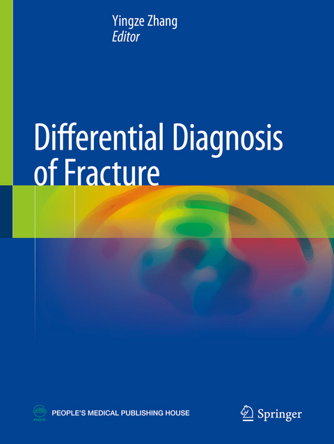
This book covers diagnostic images of common and rare fractures for nearly every part of the human body, based on a large number of clinical cases. The highlight of this book is that both of three-dimensional X-ray images and CT/MRI images of thousands of fracture cases are presented for comparison and further discussion, according to the framework of AO classification.
The first chapter gives a general introduction of various diagnostic imaging techniques for fractures, with attention to their advantages and disadvantages. The following chapters present detailed radiological images of upper extremity fractures, lower extremity fractures, axial skeleton fractures, and epiphyseal lesions. It helps readers to recognize the difference between various diagnostic techniques, and to select optimal imaging techniques. With the illustrative figures, this book is a valuable tool to orthopaedist, radiologists, trauma surgeons, emergency room doctors, professional clinical staff, and medical students.
Yingze Zhang is a Professor and Director of the orthopaedic surgery department, the Third Hospital of Hebei Medical University, Shijiazhuang, China. Professor Zhang is an academician of the Chinese Academy of Engineering (Division of Medicine and Health), and current President of Chinese Orthopedic Association (COA). He is also the author of Clinical Classification in Orthopaedics Trauma, Springer, 2018.
General Introduction.- Shoulder Joint Fracture.- Elbow Fracture.- Wrist Fracture.- Hand Fracture.- Upper Extremity Shaft Fracture.- Upper Extremity Pathological Fracture.- Hip Fracture.- Knee Joint Fracture.- Ankle Fracture.- Foot Fracture (Including Sesamoid Fracture).- Lower Extremity Shaft Fracture.- Lower Extremity Pathological Fracture.- Cervical Fracture.- Chest and Lumbar Fracture.- Sacrum and Coccyx Fracture.- Pelvic Ring Fracture.- Axial Bone Pathological Fracture.- Cartilage and Osteochondral Injury.- Occult Fracture and Contusion.- Epiphysis Injury.
| Erscheinungsdatum | 06.12.2021 |
|---|---|
| Zusatzinfo | 580 Illustrations, color; 24 Illustrations, black and white; XII, 905 p. 604 illus., 580 illus. in color. |
| Verlagsort | Singapore |
| Sprache | englisch |
| Maße | 210 x 279 mm |
| Themenwelt | Medizinische Fachgebiete ► Chirurgie ► Unfallchirurgie / Orthopädie |
| Medizinische Fachgebiete ► Radiologie / Bildgebende Verfahren ► Radiologie | |
| Schlagworte | Axial fracture • Cartilage and epiphysis injury • diagnostic radiology • Fracture imaging • Lower extremity fracture • Upper extremity fracture |
| ISBN-10 | 981-13-8341-3 / 9811383413 |
| ISBN-13 | 978-981-13-8341-0 / 9789811383410 |
| Zustand | Neuware |
| Haben Sie eine Frage zum Produkt? |
aus dem Bereich


