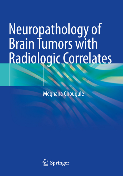
Neuropathology of Brain Tumors with Radiologic Correlates
Springer Verlag, Singapore
978-981-15-7128-2 (ISBN)
This book uses concise text and a consistent point-wise format that makes reading and reviewing easy. The radiological and pathological correlates of brain and spine tumors serve as a ready-reference resource for residents, surgical and neuropathologists, neuroradiologists, neurosurgeons, neuro-oncologists and research scientists.
Dr Meghana Chougule holds an MD in Pathology with a gold medal from Shivaji University. She is the chief consultant at Shanti Pathology Laboratory, Maharashtra. She was also an Associate Professor of Pathology at D.Y. Patil Medical College, Maharashtra. She also gained neuropathology experience at NIMHANS (National Institute of Mental Health and Neurosciences), Bangalore; TATA Memorial Hospital, Mumbai; Bombay Hospital; and Memorial Sloan Kettering Cancer Center (MSKCC), New York. Her areas of interest include neuropathology and immunohistochemistry. She has been a speaker at various conferences and has published papers in peer-reviewed international journals. She is a member of the Neuropathology Society of India and Indian Association of Pathology and Microbiology.
Squash staining - new procedure.- Diffuse astrocytic and oligodendroglial tumors.- Other astrocytic tumors.- Ependymal tumors.- Neuronal and glio-neuronal tumors.- Embryonal tumors.- Meningeal tumors.- Mesenchymal, non-menigothelial tumors.- Tumors of sellar region.- Germ cell tumors.- Choroid plexus tumors.- Cranial and paraspinal tumors.- Lymphoma.- Plasmacytoma.- Metastatic tumors.- Benign cysts of CNS.- Fungus on Squash.- Pituitary adenoma.- List of spinal tumors, Intra-axial/extra-axial, intra-dural and extra-dural tumors.
| Erscheinungsdatum | 25.10.2021 |
|---|---|
| Zusatzinfo | 412 Illustrations, color; 81 Illustrations, black and white; XV, 361 p. 493 illus., 412 illus. in color. |
| Verlagsort | Singapore |
| Sprache | englisch |
| Maße | 178 x 254 mm |
| Themenwelt | Medizinische Fachgebiete ► Chirurgie ► Neurochirurgie |
| Medizinische Fachgebiete ► Radiologie / Bildgebende Verfahren ► Radiologie | |
| Studium ► 2. Studienabschnitt (Klinik) ► Pathologie | |
| Schlagworte | Germ Cell Tumors • neuroimaging • Neuronal and glio-neuronal tumors • Pituitary adenoma • Squash staining |
| ISBN-10 | 981-15-7128-7 / 9811571287 |
| ISBN-13 | 978-981-15-7128-2 / 9789811571282 |
| Zustand | Neuware |
| Informationen gemäß Produktsicherheitsverordnung (GPSR) | |
| Haben Sie eine Frage zum Produkt? |
aus dem Bereich


