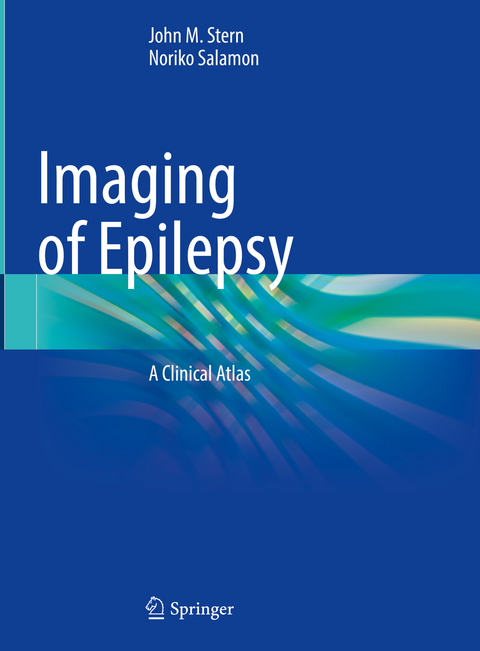
Imaging of Epilepsy
Springer International Publishing (Verlag)
978-3-030-86671-6 (ISBN)
The atlas is divided into sections according to general clinical categories with each category including a collection of clinical examples that span the category. Each example includes images across the relevant imaging modalities that relate to one patient, whose history accompanies the images. This case-based organization with clinical history and multiple images offers a complete visual understanding of the imaging findings and the corresponding relationship of each finding to the clinical presentation, treatment, and outcome.
Images for the book are from the UCLA Seizure Disorder Center, which is a referral center that serves a large outpatient epilepsy patient population and performs approximately 500 inpatient epilepsy evaluations annually.
Comprehensive and richly illustrated, this book will serve as a convenient resource in neurologic andradiologic practice, and useful for board exam review.
lt;p>Dr. John M. Stern is Professor and Director of the Epilepsy Clinical Program in the Department of Neurology at the Geffen School of Medicine, UCLA.
Dr. Noriko Salamon is Professor and Section Chief of Neuroradiology in the Department of Radiology at the Geffen School of Medicine, UCLA.
Hippocampal Sclerosis.- Focal cortical dysplasias.- Heterotopias.- Polymicrogyria.- Lissencephaly.- Schizencephaly.- Hemimegenceaphly.- Porencephaly.- Trauma.- Non-penetrating trauma.- Penetrating trauma.- Herpes encephalitis.- Neurocysticercosis.- Limbic encephalitis.- Hashimoto's encephalitis.- Rasmussen encephalitis.- Cavernous malformation.- Arteriovenous malformation.- Ischemic infarction.- Sturge-Weber syndrome.- Astrocytoma.- DNET.- Ganglioglioma.- Oligodendroglioma.- Normal hippocampal atrophy.- Atypical gyral anatomy.- Developmental venous anomaly.- Splenium signal changes.- Cerebellar atrophy.- Subarachnoid cysts.- Intra-parenchymal cysts.- Responsive neurostimulation.- Deep brain stimulation.- Anterior temporal lobe resections.- Focal cortical resection.- Hemispherectomy.- Corpus callosotomy.- Multiple sub-pial transsection.- Stereotactic thermo-ablation.- Gamma radiation.
| Erscheinungsdatum | 05.01.2022 |
|---|---|
| Zusatzinfo | XIII, 389 p. 776 illus., 208 illus. in color. |
| Verlagsort | Cham |
| Sprache | englisch |
| Maße | 210 x 279 mm |
| Gewicht | 1327 g |
| Themenwelt | Medizin / Pharmazie ► Allgemeines / Lexika |
| Medizin / Pharmazie ► Medizinische Fachgebiete ► Neurologie | |
| Medizin / Pharmazie ► Medizinische Fachgebiete ► Radiologie / Bildgebende Verfahren | |
| Schlagworte | Cortical development • Epilepsy • MRI • neuroimaging • PET • Seizures |
| ISBN-10 | 3-030-86671-8 / 3030866718 |
| ISBN-13 | 978-3-030-86671-6 / 9783030866716 |
| Zustand | Neuware |
| Haben Sie eine Frage zum Produkt? |
aus dem Bereich


