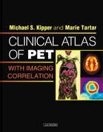
Clinical Atlas of PET with Imaging Correlation
W B Saunders Co Ltd (Verlag)
978-0-7216-3926-0 (ISBN)
- Titel ist leider vergriffen;
keine Neuauflage - Artikel merken
This user-friendly atlas demonstrates all of the major clinical applications of PET scanning. Its case-based approach with teaching points, pitfalls, and mimics presents all of the material in a concise and practical manner. And, correlative cross-sectional images illustrate the clinical features depicted in PET findings.
Chapter 1:The Basics of PET Chapter 2:Approach to PET Image Interpretation, Normal Variants, and Benign Processes Chapter 3:Lung Cancer Case 1: SPN, positive, no other disease sites (proven adenocarcinoma). Case 2: SPN, positive (proven adenocarcinoma). Case 3: SPN, positive (proven recurrent squamous cell carcinoma). Case 4: SPN, positive (proven large cell carcinoma). Case 5: SPN, positive (proven squamous cell carcinoma). Case 6: SPN, negative, tuberculoma. Case 7: SPN, negative, aspergillosis. Case 8: SPN, negative, cocci granuloma. Case 9: SPN-positive, with hilar and/or mediastinal disease (proven large cell carcinoma). Case 10: SPN, positive, with false positive mediastinal disease (proven squamous cell carcinoma). Case 11: SPN-positive, with hilar and/or mediastinal disease (advanced aquamous cell carcinoma). Case 12: Lung cancer staging, primary positive, no other disease (large cell undifferentiated carcinoma). Case 13: Lung cancer staging, primary positive (proven adenocarcinoma). Case 14: Lung cancer staging, primary only positive (NSCLC), coexisting infiltrative lung disease. Case 15: Lung cancer staging, primary site only positive (proven squamous cell carcinoma). Case 16: Lung cancer staging, with subtle mediastinal involvement (squamous cell carcinoma). Case 17: Non-small cell lung cancer, with mediastinal involvement. Case 18: Lung cancer staging, with mediastinal disease (large cell carcinoma). Case 19: Lung cancer staging, with bone metastasis (adenocarcinoma). Case 20: Lung cancer staging, with bone metastases (large cell malignancy). Case 21: Lung cancer staging, Stage IV non-small cell lung cancer, with adrenal metastasis. Case 22: Lung cancer, restaging post-treatment, with adrenal metastasis (large cell carcinoma). Case 23: Squamous cell carcinoma, Stage IV, with axillary node involvement. Case 24: Lung cancer (SCC), with chest wall invasion. Case 25: Known lung cancer for staging, extensive disease (squamous cell carcinoma). Case 26: Lung cancer staging, extensive disease (NSCLC). Case 27: Lung cancer staging, small cell carcinoma. Case 28: Lung cancer staging, localized pleural involvement (NSCLC and adenocarcinoma). Case 29: Lung cancer staging, pleural adenocarcinoma. Case 30: Non-small cell lung carcinoma, metastatic to pleura. Case 31: Lung cancer restaging, treatment assessment (SCC). Case 32: Lung cancer restaging, radiation therapy effects (SCC). Case 33: Bilateral lung cancers. Case 34: Breast carcinoma pulmonary metastases mimicking synchronous lung cancers. Case 35: Bilateral lung cancers. Case 36: Pitfall: Bronchoalveolar cell carcinoma. Case 37: Pitfall: Bronchoalveolar cell carcinoma. Case 38: Pitfall: Bronchoalveolar cell carcinoma. Case 39: Pitfall: Lung cancer false positive, pulmonary infarct. Case 40: Pitfall: lung cancer false positive, active tuberculosis. Case 41: Pitfall: lung cancer false positive, sarcoidosis. Case 42: Pitfall: Lung cancer, size limitation. Chapter 4: Lymphoma Case 1: Lymphoma, limited stage disease, staging. Case 2: Lymphoma, staging, isolated neck disease. Case 3: Lymphoma, staging and treatment assessment, pulmonary parenchymal involvement. Case 4: Unexpected PET identification of limited stage visceral lymphoma. Case 5: Bulky thoracic NSHD, initial staging and residual mass treatment assessment. Case 6: Thoracic HD, initial staging and treatment assessment. Case 7: Thoracic and abdominal HD: initial staging, treatment assessment, marrow activation. Case 8: Lymphoma, nodal & visceral involvement, staging & treatment assessment, marrow activation. Case 9: Recurrent lymphoma, bone marrow involvement. Case 10: Recurrent pelvic NHL, radiation therapy follow-up. Case 11: Mantle cell lymphoma, radiation response. Case 12: Mediastinal recurrent lymphoma, response to chemotherapy. Case 13: Mesenteric NHL, residual mass assessment. Case 14: Lymphoma, staging & treatment assessment, head & neck presentation. Case 15: Recurrent lymphoma, pediatric patient. Case 16: Lymphoma follow-up, thymic rebound. Chapter 5:Melanoma Case 1: Staging, persistent disease at operative site Case 2: Initial and re-staging, disease progression despite chemotherapy. Case 3: Staging, unexpected additional disease site. Case 4: Staging, multiple unexpected additional disease sites. Case 5: Melanoma, restaging, solitary recurrent disease site (axilla). Case 6: Restaging, recurrent facial disease. Case 7: Restaging, progressive facial recurrences. Case 8: Melanoma, restaging, lung and brain metastases. Case 9: Restaging; lung, hilar, liver and osseous metastases. Case 10: Restaging, extensive disease, treatment assessment. Case 11: Restaging, advanced disease (carcinomatosis). Chapter 6:Colorectal Cancer Case 1: Initial staging, no additional disease site (rectal cancer). Case 2: Initial staging, involved adjacent lymph node. Case 3: Initial staging, distant metastases. Case 4: Restaging, solitary liver metastasis. Case 5: Restaging, recurrent hepatic metastasis. Case 6: Restaging, solitary liver metastasis. Case 7: Restaging, retroperitoneal nodal recurrence, assessment of post-treatment pre-sacral mass. Case 8: Restaging, pelvic recurrences. Case 9: Restaging, pre-sacral recurrence. Case 10: Restaging, pulmonary parenchymal and pleural recurrences. Case 11: Recurrent colorectal carcinoma, rising CEA, bone metastases. Case 12: Colon cancer, liver metastasis, treatment effect (RF ablation). Case 13: Liver metastasis, assessment of RF ablation efficacy. Case 14: Liver metastases, surgical and interventional treatment effects. Case 15: Liver metastases, chemotherapy efficacy. Case 16: Pitfall: Recurrence, sub-centimeter lung metastases. Case 17: Pitfall: Coexisting benign disease (viral axillary adenitis). Chapter 7:Other Gastrointestinal Cancers Case 1: Proximal esophageal squamous cell carcinoma. Case 2: Distal esophageal adenocarcinoma, with gastro-hepatic nodal involvement. Case 3: Esophageal carcinoma, superior mediastinal paraesophageal nodal involvement. Case 4: Distal esophageal SCC. Case 5: Distal esophageal adenocarcinoma, with gastric cardia extension and paragastric nodal involvement. Case 6: Gastric carcinoma, with retroperitoneal nodal metastases. Case 7: Gastric carcinoma, with peritoneal carcinomatosis. Case 8: Pancreatic adenocarcinoma. Case 9: Locally recurrent pancreatic carcinoma. Case 10: Recurrent pancreatic carcinoma, metastatic to liver and brain. Case 11: Ampullary adenocarcinoma, with local nodal involvement. Case 12: Cholangiocarcinoma, with liver metastasis. Case 13: Recurrent cholangiocarcinoma, drop metastasis. Case 14: Suspected residual gallbladder carcinoma. Chapter 8:Head and Neck Cancer Case 1: Normal head and neck anatomy example. Case 2: Glottic squamous cell carcinoma, initial diagnosis. Case 3: Initial staging, BOT squamous cell carcinoma, with nodal metastasis at presentation. Case 4: Locally advanced base of tongue squamous cell carcinoma, with bilateral necrotic lymph node metastases. Case 5: Metastatic squamous cell carcinoma, cervical lymph node presentation, primary lesion search. Case 6: Hard palate squamous cell carcinoma, radiation therapy planning. Case 7: Recurrent maxillary non-small cell malignancy. Case 8: Recurrent and progressive squamous cell carcinoma. Case 9: Nasopharyngeal squamous cell carcinoma, with bone metastases. Case 10: Squamous cell carcinoma, metastatic to thoracic spine, incipient cord compression presentation. Chapter 9:Breast Cancer Case 1: Focal breast activity due to an unsuspected breast cancer. Case 2: Breast cancer, initial staging, axillary nodal presentation, primary search and internal mammary adenopathy. Case 3: Breast cancer restaging, normal post-lumpectomy and radiation breast findings. Case 4: Post surgical biopsy scar, post lumpectomy for infiltrating ductal carcinoma. Case 5: Treated inflammatory breast cancer. Case 6: Recurrent inflammatory breast cancer. Case 7: Breast cancer restaging, in situ neoadjuvantly treated infiltrating lobular carcinoma, with diffuse blastic bone metastases. Case 8: Breast cancer restaging, bone metastases. Case 9: Breast cancer restaging, active bone metastases, treated liver metastases. Case 10: Recurrent breast cancer, with extensive liver metatases. Case 11: Breast cancer restaging, solitary liver metatasis. Case 12: Recurrent breast cancer, with chest wall and lung parenchymal disease. Case 13: Breast cancer restaging, axillary and chest wall involvement and bone metastases. Case 14: Breast cancer restaging, extensive local and nodal recurrence. Case 15: Breast cancer restaging, local and nodal recurrences in axilla, supraclavicular neck and mediastinum. Case 16: Breast cancer restaging; mediastinal, neck and supraclavicular nodal recurrences. Case 17: Breast cancer restaging, hilar nodal involvement, progression to liver and bone metastases. Case 18: Restaging, thoracic (nodal and pulmonary parenchymal) metastases. Case 19: Breast Cancer restaging, assessment of chemotherapy efficacy, mediastinal and bone metastases. Chapter 10:Miscellaneous Tumors Case 1: Recurrent thyroid carcinoma, lungs and neck. Case 2: Recurrent thyroid carcinoma, isolated neck lymph node. Case 3: Recurrent thyroid carcinoma to neck, low metabolic rate. Case 4: Pitfall: Suspected recurrent thyroid carcinoma to mediastinum, false positive (thymus) Case 5: In situ primary, presenting with pleural metastases Case 6: Sarcomatoid renal cell carcinoma, with retroperitoneal metastases Case 7: In situ primary, with IVC tumor extension Case 8: Renal cell carcinoma, lung metastasis. Case 9: Recurrent renal cell carcinoma, hilar and vertebral metastases Case 10: Locally recurrent clear cell renal cell carcinoma, with lung metastases Case 11: Widely metastatic testicular carcinoma, response to chemotherapy Case 12: Suspected recurrence, disseminated sarcoidosis Case 13: In situ primary transitional cell carcinoma Case 14: Widely metastatic transitional cell carcinoma Case 15: Widely metastatic prostate cancer, dedifferentiated Case 16: Benign adrenal hemangioendothelioma Case 17: Recurrent ovarian carcinoma, response to chemotherapy Case 18: Recurrent ovarian carcinoma, with parathyroid adenoma Case 19: Local recurrence of cervical carcinoma, pararectal region with synchronous lung colon carcinoma metastasis Case 20: Widely metastatic cervical carcinoma (brain, porta hepatic, supraclavicular and presacral) Case 21: Uterine corpus carcinoma, with vaginal metastasis Case 22: Unsuspected recurrent leiomyosarcoma to multiple muscles Case 23: Synovial sarcoma, metastatic to lung Case 24: Recurrent intraabdominal leiomyosarcoma Case 25: Recurrent thoracic liposarcoma Case 26: Ewing's sarcoma follow-up Case 27: Residual leiomyosarcoma, post operative evaluation for residual disease Case 28: Malignant thymoma, pleural recurrence Case 29: Pitfall: Inflammatory reaction, suspected anterior mediastinal thymoma Case 30: Multiple myeloma, initial staging Case 31: Multiple discrete lesions, known disease follow-up Case 32: Multiple myeloma, diffusely infiltrative, poorly demonstrated on PET Case 33: Kaposi's sarcoma (non-AIDS) Case 34: Bowen's (multiple squamous cell carcinoma) Chapter 11:Neurologic PET Applications Case 1: Normal brain PET: guidelines for image interpretation. Case 2: Recurrent glioblastoma multiforme (differentiation from radiation necrosis). Case 3: Lung cancer metastasis, gamma knife follow-up. Case 4: Low-grade oligodendroglioma, initial diagnosis. Case 5: Oligodendroglioma, tumor differential diagnosis. Case 6: Low-grade glioma, transformation. Case 7: MCA infarct. Case 8: Radiation necrosis, residual oligodendroglioma. Case 9: Radiation necrosis, s/p scalp melanoma therapy. Case 10: Bitemporal radiation necrosis, s/p nasopharyngeal carcinoma therapy. Case 11: Temporal radiation necrosis, s/p pre-auricular basal cell carcinoma therapy, abnormal brain SPECT. Case 12: Alzheimer's. Case 13: Alzheimer's. Case 14: Pick's (frontal lobe dementia). Case 15: Primary cerebellar degeneration. Case 16: Temporal lobe hypometabolism Chapter 12:Cardiac PET Applications Case 1: Myocardial viability study: Normal example Case 2: Myocardial viability study: Patient with non-Q wave MI and CHF, with abnormal thallium viability study Case 3: Myocardial viability study: Patient with known CAD, post MI and PTCA, with recurrent angina and abnormal SPECT Case 4: Myocardial viability study: Patient with chronic CHF post MI, being considered for percutaneous revascularization for fatigue and chest pain Case 5: Myocardial viability study: Nonsurgical candidate patient with recurrent symptoms, with abnormal SPECT, being considered for repeat percutaneous intervention Case 6: Myocardial viability study: Diabetic patient with multi-vessel CAD and ischemic cardiomyopathy, being considered for CABG revascularization Case 7: Myocardial viability study: Patient with prior MI, CABG, PTCA and ischemic cardiomyopathy, with matched perfusion/metabolism defects Case 8: Myocardial viability study: Patient with long-standing CAD, post multiple revascularization procedures, with persistent angina and dypnea and recurrent disease by angiography Case 9: Myocardial viability study: Diabetic, vasculopathic, high surgical risk patient, with abnormal SPECT and poor LV function, being considered for CABG
| Erscheint lt. Verlag | 13.1.2004 |
|---|---|
| Zusatzinfo | Approx. 1100 illustrations |
| Verlagsort | London |
| Sprache | englisch |
| Maße | 216 x 276 mm |
| Gewicht | 1650 g |
| Themenwelt | Medizinische Fachgebiete ► Radiologie / Bildgebende Verfahren ► Computertomographie |
| ISBN-10 | 0-7216-3926-7 / 0721639267 |
| ISBN-13 | 978-0-7216-3926-0 / 9780721639260 |
| Zustand | Neuware |
| Haben Sie eine Frage zum Produkt? |
aus dem Bereich


