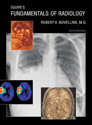
Squire's Fundamentals of Radiology
Harvard University Press (Verlag)
978-0-674-01279-0 (ISBN)
- Titel ist leider vergriffen;
keine Neuauflage - Artikel merken
In the past five years, the development of new imaging technologies that make possible faster and more accurate diagnoses has significantly improved the imaging of disease and injury. This new edition of Squire’s Fundamentals of Radiology describes and illustrates these new techniques to prepare medical students and other radiology learners to provide the most optimal and up-to-date imaging management for their patients. Not only are new diagnostic techniques outlined, such as the multidetector computed tomography diagnosis of pulmonary embolism and the diffusion-weighted magnetic-resonance imaging of stroke, but hundreds of new diagnostic images have been included to illustrate the radiological characteristics of common diseases with state-of-the-art computed radiography, ultrasound, multidetector computed tomography, and magnetic-resonance images. The text has been completely reviewed and updated to present the latest and best strategies in diagnostic imaging.
New interventional radiology procedures have been added, including vertebroplasty, a percutaneous injection treatment of painful spinal compression fractures; uterine artery embolization, a surgical alternative to hysterectomy in women with painful or bleeding uterine fibroids; and radiofrequency ablation, a percutaneous technique for treating unresectable tumors in the liver and other organs with probes that superheat and thus destroy cancer cells.
A new chapter on advances in diagnostic imaging describes many cutting-edge imaging technologies, such as three-dimensional and digital imaging, functional magnetic-resonance imaging, PET–CT (positron emission tomography combined with computed tomography), cardiac calcium CT scoring, multidetector gated cardiac CT, and molecular imaging.
Robert A. Novelline, M.D., is Professor Emeritus of Radiology at Harvard Medical School and Director Emeritus of both the Harvard Medical School Core and Advanced Radiology Student Clerkships at Massachusetts General Hospital.
1. Basic Concepts Radiodensity as a Function of Thickness Radiodensity as a Function of Composition, with Thickness Kept Constant How Roentgen Shadows Instruct You about Form Radiographs as Summation Shadowgrams 2. The Imaging Techniques Thinking Three-Dimensionally about Plain Films The Routine Posteroanterior (PA) Film PA and AP Chest Films Compared The Lateral Chest Film The Lordotic View Conventional Tomography Radiographs of Coronal Slices of a Frozen Cadaver Conventional Tomograms of the Living Patient in the Coronal Plane Fluoroscopy Angiography Computed Tomography Reformatted CT Images in Coronal, Sagittal, and Other Planes and Three-Dimensional CT CT Angiography Ultrasound Magnetic-Resonance Imaging Radioisotope Scanning 3. Normal Radiological Anatomy 4. How to Study the Chest Projection The Rib Cage Confusing Shadows Produced by Rotation The Importance of Exposure Soft Tissues 5. The Lung The Normal Lung Variations in Pulmonary Vascularity The Pulmonary Microcirculation Variations in the Pulmonary Microcirculation Solitary and Disseminated Lesions in the Lung Air-Space and Interstitial Disease The Importance of Clinical Findings High-Resolution CT of the Lung 6. Lung Consolidations and Pulmonary Nodules Consolidation of a Whole Lung Consolidation of One Lobe Consolidation of Only a Part of One Lobe Solitary and Multiple Pulmonary Nodules 7. The Diaphragm, the Pleural Space, and Pulmonary Embolism Pleural Effusion Pneumothorax Pulmonary Embolic Disease Radioisotope Perfusion and Ventilation Lung Scans Pulmonary Embolism CT 8. Lung Overexpansion, Lung Collapse, and Mediastinal Shift Emphysema Normal Mediastinal Position Mediastinal Shift 9. The Mediastinum Mediastinal Compartments and Masses Arising within Them Anterior Mediastinal Masses Anterior and Middle Mediastinal Masses Posterior Mediastinal Masses 10. The Heart Measurement of Heart Size Factors Limiting Measurement of Heart Size Examples of Apparent Abnormality in Heart Size and Difficulties in Measuring Interpretation of the Measurably Enlarged Heart Shadow Enlargement of the Left or Right Ventricle The Heart in Failure Variations in Pulmonary Blood Flow Cardiac Calcification The Anatomy of the Heart Surface Identifying Right and Left Anterior Oblique Views The Anatomy of the Heart Interior Coronary Arteriography Classic Changes in Shape with Chamber Enlargement Nuclear Cardiac Imaging MR Images of the Heart Cardiac CT 11. How to Study the Abdomen The Plain Film Radiograph Identifying Parts of the Gastrointestinal Tract Identifying Fat Planes Identifying Various Kinds of Abnormal Densities Systematic Study of the Plain Film CT of the Abdomen Ultrasound of the Abdomen 12. Bowel Gas Patterns, Free Fluid, and Free Air The Distended Stomach The Distended Colon The Distended Small Bowel Differentiating Large-Bowel and Small-Bowel Obstruction from Paralytic Ileus Free Peritoneal Fluid Free Peritoneal Air 13. Contrast Study and CT of the Gastrointestinal Tract Principles of Barium Work Normal Variation versus a Constant Filling Defect The Components of the Upper GI Series Rigidity of the Wall Filling Defects and Intraluminal Masses in the Stomach and Small Bowel Gastric and Duodenal Ulcers The Barium Enema Filling Defects and Intraluminal Masses in the Colon CT of the Gastrointestinal Tract CT of the Thickened Bowel Wall CT of Diverticular Disease CT of Appendicitis CT of Bowel Obstruction CT of Bowel Ischemia 14. The Abdominal Organs The Liver Liver Metastases Primary Tumors of the Liver Hepatic Cysts and Abscesses Liver Trauma Cirrhosis, Splenomegaly, and Ascites Splenic Trauma Cholelithiasis and Cholecystitis Obstruction of the Biliary Tree The Pancreas Pancreatic Tumors Pancreatitis and Pancreatic Abscesses Pancreatic Trauma The Urinary Tract Obstructive Uropathy Cystic Disease of the Kidneys Urinary Tract Infection Renal Tumors Intravenous Contrast Materials Renal Trauma The Urinary Bladder The Adrenal Glands 15. The Musculoskeletal System How to Study Radiographs of Bones Requesting Films of Bones Fractures Fracture Clinic Dislocations and Subluxations Osteomyelitis Arthritis Osteonecrosis Microscopic Bone Structure and Maintenance The Development of Metabolic Bone Disease Osteoporosis of the Spine Spine Fractures Osteomyelitis of the Spine Metastatic Bone Tumors Primary Bone Tumors Musculoskeletal MR Imaging 16. Men, Women, and Children The Female Breast The Female Pelvis Gynecological Conditions Obstetrical Imaging Ectopic Pregnancy Placenta Previa Placental Abruption The Scrotum The Prostate The Male Urethra Croup and Epiglottitis Pneumonia, Bronchiolitis, and Bronchitis Cystic Fibrosis Hypertrophic Pyloric Stenosis Ileocolic Intussusception Hirschsprung's Disease Abdominal Masses in Infants and Children Normal Pediatric Bones Fractures in Children Child Abuse Pediatric Cranial Ultrasound 17. The Vascular System Conventional Arteriography Digital Subtraction Angiography Conventional Venography Ultrasound and Color Doppler Ultrasound MR Angiography CT Angiography Arterial Anatomy Aortic Aneurysm Aortic Dissection Traumatic Aortic Injury Atherosclerotic Arterial Occlusive Disease Renovascular Hypertension Venous Anatomy Obstruction of the Superior Vena Cava Disorders of the Inferior Vena Cava Deep Venous Thrombosis of the Lower Extremities 18. The Central Nervous System Imaging Techniques CT Anatomy of the Normal Brain MR Anatomy of the Normal Brain CT and MR Compared Hydrocephalus, Brain Atrophy, and Intracranial Hemorrhage Normal Cerebral Arteriography Head Trauma Cerebrovascular Disease and Stroke Brain Tumors Cerebral Aneurysm and Arteriovenous Malformation The Face Low Back Pain and Lumbar Disc Syndrome Spinal Tumors 19. Interventional Radiology Percutaneous Transluminal Angioplasty Transcatheter Embolization Angiographic Diagnosis and Control of Acute Gastrointestinal Hemorrhage Inferior Vena Cava Filters Image-Guided Venous Access Percutaneous Aspiration Needle Biopsy of the Thorax Percutaneous Aspiration Needle Biopsy of the Abdomen Percutaneous Abscess Drainage Percutaneous Gastrostomy and Jejunostomy Radiofrequency Ablation Percutaneous Cholecystotomy Radiological Management of Urinary Tract Obstruction Uterine Fibroid Embolization Interventional Neuroradiology Vertebroplasty 20. The Latest in Diagnostic Imaging Virtual Colonoscopy and Virtual Bronchoscopy Coronary Calcium Scoring Coronary CT Angiography Three-Dimensional Ultrasound Fusion Imaging with PET-CT Functional MR Imaging Molecular Imaging Answers to Unknowns Acknowledgments Index
| Erscheint lt. Verlag | 2.4.2004 |
|---|---|
| Zusatzinfo | 1300 halftones, 80 line illustrations, 20 color illustrations |
| Verlagsort | Cambridge, Mass |
| Sprache | englisch |
| Maße | 216 x 279 mm |
| Gewicht | 2422 g |
| Themenwelt | Medizinische Fachgebiete ► Radiologie / Bildgebende Verfahren ► Kernspintomographie (MRT) |
| Medizinische Fachgebiete ► Radiologie / Bildgebende Verfahren ► Radiologie | |
| Medizinische Fachgebiete ► Radiologie / Bildgebende Verfahren ► Sonographie / Echokardiographie | |
| ISBN-10 | 0-674-01279-8 / 0674012798 |
| ISBN-13 | 978-0-674-01279-0 / 9780674012790 |
| Zustand | Neuware |
| Haben Sie eine Frage zum Produkt? |
aus dem Bereich


