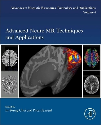
Advanced Neuro MR Techniques and Applications
Academic Press Inc (Verlag)
978-0-12-822479-3 (ISBN)
Trainees such as postdoctoral fellows, PhD and MD/PhD students, residents and fellows using or considering the use of neuro MR technologies will also be interested in this book.
In-Young Choi, Ph.D. is Professor in the Department of Neurology at the University of Kansas Medical Center. She is also a faculty member with the Department of Molecular & Integrative Physiology and Hoglund Brain Imaging Center, and is affiliated with the Bioengineering Program at the University of Kansas. Dr. Choi received her Ph.D. in Biophysical Sciences and Medical Physics from the University of Minnesota and was trained in advanced magnetic resonance imaging / spectroscopy techniques and neurobiology at the Center for Magnetic Resonance Research at the University of Minnesota. Dr. Choi’s research focuses largely on the identification of quantitative, objective biomarkers of the pathologic mechanisms underlying disease status and progression in a variety of neurological conditions to characterize metabolic, morphological and functional pathophysiology of the disease, and to guide and accelerate the development of new treatment strategies. Dr. Choi works on the development of advanced noninvasive in vivo magnetic resonance imaging and spectroscopy techniques for interdisciplinary translational research, encompassing biomedical imaging, neurology, neurobiology, and neurochemistry. She is a member of the editorial board of Frontiers series and Neurochemical Research, and editor/guest editor of Advances in Neurobiology, NMR in Biomedicine, Neurochemical Research and Magnetic Resonance in Medicine. Professor Jezzard’s FMRIB Physics Group, part of the Wellcome Centre for Integrative Neiroimaging, develops novel physiological MRI methods for the study of healthy and diseased brain. He trained in physics in the UK, before joining the National Institutes of Health, USA, between 1991-1998. Since 1998 he has been based at the University of Oxford, UK. He is particularly interested in techniques for mapping the macroscopic and microscopic neurovasculature, and collaborates closely with various clinical groups, in particular through the Oxford Acute Vascular Imaging Centre (AVIC), on the development of rapid imaging approaches to aid in the diagnosis and treatment of stroke and cerebrovascular disease. A second thread of his research aims to advance ultra-high field imaging. This research combines novel imaging hardware, including parallel RF transmission, with state-of-the-art acquisition techniques. He holds leadership roles in several imaging centres within Oxford, and has been active in the International Society for Magnetic Resonance in Medicine in a range of capacities, including as President from 2013-2014.
Part 1: Fast and Robust Imaging 1. Recommendations for Neuro MRI Acquisition Strategies 2. Advanced Reconstruction Methods for Fast MRI 3. Simultaneous Multi-Slice MRI 4. Motion Artifacts and Correction in Neuro MRI
Part 2: Classical and Deep Learning Approaches to Neuro Image Analysis 5. Statistical Approaches to Neuroimaging Analysis 6. Image Registration 7. Image Segmentation
Part 3: Diffusion MRI 8. Diffusion MRI Acquisition and Reconstruction 9. Diffusion MRI Artifact Correction 10. Diffusion MRI Analysis Methods 11. Diffusion MRI as a Probe of Tissue Microstructure
Part 4: Perfusion MRI 12. Non-Contrast Agent Perfusion MRI Methods 13. Contrast Agent-Based Perfusion MRI Methods 14. Perfusion MRI: Clinical Perspectives
Part 5: Functional MRI 15. Functional MRI Principles and Acquisition Strategies 16. Functional MRI Analysis 17. Neuroscience Applications of Functional MRI 18. Clinical Applications of Functional MRI
Part 6: The Brain Connectome 19. The Diffusion MRI Connectome 20. Functional MRI Connectivity 21. Applications of MRI Connectomics
Part 7: Susceptibility MRI 22. Principles of Susceptibility-Weighted MRI 23. Applications of Susceptibility-Weighted Imaging and Mapping
Part 8: Magnetization Transfer Approaches 24. Magnetization Transfer Contrast MRI 25. Chemical Exchange Saturation Transfer (CEST) MRI as a Tunable Relaxation Phenomenon 26. Clinical Application of Magnetization Transfer Imaging
Part 9: Quantitative Relaxometry and Parameter Mapping 27. Quantitative Relaxometry Mapping 28. MR Fingerprinting: Concepts, Implementation and Applications 29. Quantitative Multi-Parametric MRI Measurement
Part 10: Neurovascular imaging 30. Neurovascular Magnetic Resonance Angiography 31. Neurovascular Vessel Wall Imaging: New Techniques and Clinical Applications
Part 11: Advanced Magnetic Resonance Spectroscopy 32. Single Voxel Magnetic Resonance Spectroscopy: Principles and Applications 33. Magnetic Resonance Spectroscopic Imaging: Principles and Applications 34. Non-Fourier-Based Magnetic Resonance Spectroscopy
Part 12: Ultra-high Field Neuro MR Techniques 35. Benefits, Challenges and Applications of Ultra-High Field Magnetic Resonance 36. Neuroscience Applications of Ultra-High-Field Magnetic Resonance Imaging: Mesoscale Functional Imaging of the Human Brain 37. Clinical Applications of High Field Magnetic Resonance
| Erscheinungsdatum | 02.12.2021 |
|---|---|
| Reihe/Serie | Advances in Magnetic Resonance Technology and Applications |
| Zusatzinfo | 63 illustrations (48 in full color); Illustrations |
| Verlagsort | San Diego |
| Sprache | englisch |
| Maße | 191 x 235 mm |
| Gewicht | 1270 g |
| Themenwelt | Medizin / Pharmazie ► Medizinische Fachgebiete ► Neurologie |
| Medizinische Fachgebiete ► Radiologie / Bildgebende Verfahren ► Radiologie | |
| Naturwissenschaften ► Biologie ► Humanbiologie | |
| Naturwissenschaften ► Biologie ► Zoologie | |
| ISBN-10 | 0-12-822479-7 / 0128224797 |
| ISBN-13 | 978-0-12-822479-3 / 9780128224793 |
| Zustand | Neuware |
| Informationen gemäß Produktsicherheitsverordnung (GPSR) | |
| Haben Sie eine Frage zum Produkt? |
aus dem Bereich


