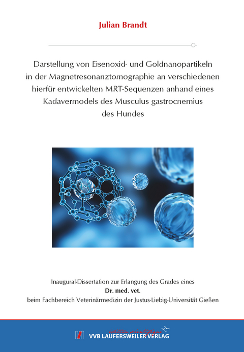
Darstellung von Eisenoxid- und Goldnanopartikeln in der Magnetresonanztomographie an verschiedenen hierfür entwickelten MRT-Sequenzen anhand eines Kadavermodels des Musculus gastrocnemius des Hundes
Seiten
2020
VVB Laufersweiler Verlag
978-3-8359-6927-8 (ISBN)
VVB Laufersweiler Verlag
978-3-8359-6927-8 (ISBN)
- Keine Verlagsinformationen verfügbar
- Artikel merken
Nanopartikel sind kleine Partikel mit einem Durchmesser von 1 bis 100nm (Soliman et al., 2015, Hühn et al., 2017). Für die Zellmarkierung spielen sie eine interessante und zukunftsrelevante Rolle, insbesondere in der fortschreitenden Entwicklung der Anwendung von Stammzellen in der regenerativen Stammzelltherapie (Kustermann et al., 2008, Arnhold & Wenisch, 2015, Kolecka et al., 2017). Die Darstellung der Nanopartikel, bzw. später der markierten Zellen, soll im Rahmen dieser Studie mit der Magnetresonanztomographie (MRT) als ein nicht invasives bildgebendes Verfahren untersucht werden (López-Lagunaa et al., 2011, Schmied, 2018). In der vorliegenden Studie werden 4nm große superparamagnetische Eisenoxid Nanopartikel (SPIO) und 100nm große goldhaltige Nanopartikel (AuNP) als MRT- Kontrastmittel in verschiedenen Konzentrationen an sieben unterschiedliche speziell zur Darstellung der Nanopartikel entwickelten MRT- Untersuchungssequenzen überprüft. Dies wird anhand eines Kadavermodels nach intramuskulärer ultraschall-gestützter Injektion der Nanopartikel am lateralen Kopf des M. gastrocnemius in Anlehnung an Muskulotendinopathien des M. gastrocnemius bei athletischen Hunderassen durchgeführt (Kaiser et al., 2016). Die hier verwendeten Konzentrationen sind um ein Vielfaches geringer als diejenigen von Vergleichsstudien zur Darstellung und Überprüfung von zellmorphologischen und funktionalen Effekten (Wen et al., 2013, Kolecka et al., 2017, Schmied, 2018).
Die Eisenoxidnanopartikel können mit dem MRT besonders kontrastreich dargestellt werden. Dabei fällt die gemessene Signalintensität mit steigender Konzentration an SPIO konzentrationsabhängig ab. Dies wird insbesondere bei Gradientechosequenzen (GRE) deutlich, da diese zur Darstellung der von SPIO verursachten Suszeptibilitätsartefakte am geeignetsten sind (Westbrook et al., 2011). Die hohe Korrelation zwischen objektiver und subjektiver Messung erlaubt eine adäquate subjektive Einschätzung seitens des Untersuchers. Für die klinische Anwendung werden folgende Sequenzen zur Darstellung der SPIO empfohlen: T1 gewichtete (w) Turbo-Spin-Echo (TSE) (ab 146,48nM) als anatomische Übersichtsaufnahme, die T2w TSE (ab 146,48nM, besser ab 219,72nM) zur Darstellung akuter pathologischer Läsionen, die T2w GRE (sagittal und transversal, ab 146,48nM) zur optimalen Darstellung der SPIO und die Short Tau Inversion Recovery (STIR) Sequenz zur Unterscheidung von akuter und chronischer Läsionen (ab 494,36nM, besser ab 741,55nM). Die protonengewichteten Aufnahmen sind für klinischen Bereich nicht relevant.
Im Gegensatz zu den Eisenoxidnanopartikeln ist der Kontrast der diamagnetischen Goldnanopartikel deutlich schwächer ausgeprägt. So zeigen die Partikel in diesem Versuchsaufbau lediglich bei der T2w GRE Sequenz einen deutlich hypointensen erkennbaren konzentrationsabhängigen Verlauf (ab 0,00336nM). Dennoch wäre es möglich, die vielseitigen anderen Eigenschaften (z.B. Oberflächenplasmonresonanz, Biosensor, CT etc.) der Goldnanopartikel zu nutzen um multimodale Kontrastmittel herzustellen, indem sie mit anderen MRT-Kontrastmittel, wie z.B. Gadolinium-Chelate, verknüpft werden (Huang et al., 2007, Alric et al., 2008, Park et al., 2010, Colombo et al., 2012).
In möglichen auf diese Arbeit aufbauenden Studien könnte untersucht werden, ob die mit SPIO markierten mesenchymalen Stammzellen in den oben genannten niedrigen Konzentrationen mittels MRT im Kadavermodell darstellbar sind. Anschließend könnte in einer weiteren in vivo Studie mittels ultraschall-gestützter Injektion die Lokalisation und der zeitliche Verbleib der markierten Stammzellen im Organismus überprüft werden. In diesem Versuchsaufbau wären auch biologische und potentiell toxikologische Fragestellungen im Organismus aufschlussreich. Schlussendlich ist es im Rahmen der regenerativen Stammzelltherapie das Ziel Patienten mit markierten Stammzellen unterstützend zu therapieren.
Das Fazit aus dieser Dissertation lautet, dass SPIO in den oben genannten Untersuchungssequenzen und Konzentrationen ausreichend Signal erzeugen, um optimal mittels MRT objektiv und subjektiv dargestellt werden zu können. Dabei begünstigt die äußerst niedrige Konzentration im Rahmen der Zellmarkierung die Zellvitalität, sodass später der gewünschte therapeutische Effekt der regenerativen Stammzelltherapie bestmöglich ausgenutzt werden kann.
Schlüsselwörter: Eisenoxidnanopartikel, Goldnanopartikel, Entwicklung von MRT-
Untersuchungssequenzen, Stammzelltherapie, Zellmarkierung, Kadavermodel, Muskulotendinopathie vom M. gastrocnemius Nanoparticles are small particles with a diameter ranged between 1 and 100nm (Soliman et al., 2015, Hühn et al., 2017). For cell labelling they play an interesting and future relevant role in the progressive development of stem cell application in regenerative stem cell therapy (Kustermann et al., 2008, Arnhold & Wenisch, 2015, Kolecka et al., 2017). For the display of nanoparticles, respectively of marked stem cells there is demand on new techniques for the evaluation of the exact localisation of the implanted particles/cells and for the therapeutic success in vivo. For this purpose, magnetic resonance imaging (MRI), as a non-invasive imaging device is investigated by this study (López-Lagunaa et al., 2011, Schmied, 2018). In this study 4nm superparamagnetic iron oxide nanoparticles (SPIO) and 100nm gold nanoparticles (AuNP) are tested as MRI contrast agents in different low concentrated dilutions on 7 different pulsesequences, which have specially been developed to image the nanoparticles. The study will be performed on a cadaver model by intramuscular ultrasound-aimed injection of contrast into the lateral head of the M. gastrocnemius according to a musculotendinopathy of athletic dog breeds (Kaiser et al., 2016). The
concentrations of nanoparticles that are used are many times lower than used in other studies for the imaging of nanoparticles or used for nanoparticles-dependant cell morphological or functional changes (López-Lagunaa et al., 2011, Schmied, 2018).
Especially the iron oxide nanoparticles can be imagined rich in contrast with MRI. There is a concentration-dependent decline of signal intensity. The higher the concentration, the lower the intensity. On the gradient-echo sequence this correlation is particularly strong because susceptibility artefacts, which are generated by SPIO, are seen superior at these kinds of sequences (Westbrook et al., 2011). Furthermore, the high correlation between objective and subjective measurement permit an authentic subjective evaluation of the examiner. We recommend the following sequences in the clinical application to imagine SPO during examination: T1 weighted (w) turbo-spin-echo (TSE) (from 146,48nM) for anatomical overview, T2w TSE (from 146,48nM, better from 219,72nM) for the imaging of acute pathological lesions, T2w GRE (sagittal and transversal, from 146,49nM) for the accurate imaging of SPIO and at least the Short Tau Inversion Recovery (STIR) sequence to differentiate between acute and chronic lesions (from 494,36nM, better from 741,55nM). The proton-density weighted sequences are less relevant for clinical purpose.
The contrast of diamagnetic gold nanoparticles is notedly weaker in contrast to the iron oxide nanoparticles. In this study the gold particles only show a clear concentration-dependant hypointense signal on the T2w GRE sequence (from 0,00336nM). The higher the concentration, the lower the signal. Though, it would still be possible to use the additional properties of gold nanoparticles (such as surface plasmon resonance, biosensorics, CT etc.) to build multimodal contrast in combination with a MRI contrast, such as gadolinium-chelate (Huang et al., 2007, Alric et al., 2008, Park et al., 2010, Colombo et al., 2012).
Further studies could deal with research that is conducted to find out whether SPIO marked stem cells in referred low concentrations are visible on MRI in the cadaver model. Furthermore, in a in vivo study it could be reviewed if ultrasound-injected stem cells are located properly and investigate their process in the organisms over time. In this study biological and potential toxicological questions could be answered as well. The purpose is to treat patients in a supportive way with marked stem cells in a regenerative therapy.
The conclusion of this dissertation is that SPIO produce enough contrast for objective and subjective magnetic resonance imaging according to the above-mentioned sequences and concentrations. At the same time the particularly low concentration of nanoparticles promotes cell vitality in case of cell labelling, so the desired therapeutic effect of regenerative stem cell therapy will be used to full capacity.
Keywords: iron oxide nanoparticle, gold nanoparticle, development of MRI-sequences
stem cell therapy, cell labelling, cadaver model, musculotendinopathy of the m. gastrocnemius
| Erscheinungsdatum | 26.01.2021 |
|---|---|
| Reihe/Serie | Edition Scientifique |
| Sprache | deutsch |
| Maße | 146 x 210 mm |
| Gewicht | 260 g |
| Themenwelt | Medizin / Pharmazie ► Allgemeines / Lexika |
| Studium ► 2. Studienabschnitt (Klinik) ► Anamnese / Körperliche Untersuchung | |
| Veterinärmedizin ► Klinische Fächer ► Bildgebende Verfahren | |
| Schlagworte | Eisenoxidnanopartikel • Gastrocnemius • Goldnanopartikel • iron oxide nanoparticle • Kadavermodel • MRI • MRT • musculotendinopathy • Muskulotendinopathie • Nanoparticle • Stammzelltherapie • Stem Cell Therapy • Untersuchungssequenzen • Zellmarkierung |
| ISBN-10 | 3-8359-6927-7 / 3835969277 |
| ISBN-13 | 978-3-8359-6927-8 / 9783835969278 |
| Zustand | Neuware |
| Haben Sie eine Frage zum Produkt? |
Mehr entdecken
aus dem Bereich
aus dem Bereich
aus Klinik und Praxis
Buch | Softcover (2023)
Urban & Fischer (Verlag)
42,00 €
Buch | Hardcover (2017)
Hogrefe (Verlag)
60,00 €
Buch | Softcover (2024)
Urban & Fischer in Elsevier (Verlag)
56,00 €


