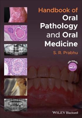
Handbook of Oral Pathology and Oral Medicine
Wiley-Blackwell (Verlag)
978-1-119-78112-7 (ISBN)
Handbook of Oral Pathology and Oral Medicine delivers a succinct overview of a range of oral diseases. The book contains up-to-date evidence-based information organized by clinical topic and supported by over 300 clinical, radiological, and microscopic images. Each chapter includes topics following universally respected curricula of oral pathology and oral medicine.
Divided into seven part, it covers core topics such as pathology of teeth, pulp, and supporting structures, pathology of jawbones, pathology of the oral mucosa, pathology of the salivary glands, clinical presentation of mucosal disease, orofacial pain, and miscellaneous topics of clinical relevance.
Written for undergraduate dental students, dental hygienists and oral health therapists, »Handbook of Oral Pathology and Oral Medicine is an ideal quick reference and is also useful to dental educators and practitioners.
S.R. Prabhu is Honorary Associate Professor of Oral Medicine at the School of Dentistry at the University of Queensland in Brisbane, Australia. He is a Fellow of Dental Faculty of all four United Kingdom and Ireland based Royal Surgical Colleges. He has taught oral pathology and oral medicine in a number of dental schools internationally, and has authored numerous book chapters, papers and reviews, and edited books.
Foreword
Preface
Acknowledgements
Standard Abbreviations
Terminology used in oral pathology and oral medicine
PART 1. PATHOLOGY OF TEETH AND SUPPORTING STRUCTURES
1. Disorders of tooth development and eruption
1. 1. Anodontia, hypodontia and oligodontia
1. 2. Hyperdontia (supernumerary teeth)
1.3. Microdontia and macrodontia
1.4. Gemination, fusion and concrescence
1.5. Taurodontism and dilaceration
1.6. Amelogenesis imperfecta
1.7. Dentinogenesis imperfecta
1.8. Dentinal dysplasia
1.9. Regional odontodysplasia
1.10. Delayed tooth eruption
1.11. Tooth impaction
1.12. Dens invaginatus and dens evaginatus
1.13. Fluorosis
1.14. Tetracycline induced discolouration of teeth
1.15. Enamel pearl,
1.16. Talon cusp
1.17. Hutchinson's incisors and mulberry molars
1.18. Tooth ankylosis
1.19. Supernumerary roots
2. Dental caries
2.1. Definition/description
2.2. Incidence/prevalence
2.3. Aetiology/risk factors/pathogenesis
2.4. Classification of caries
2.5. Clinical features
2.5.1. Primary caries
2.5.2. Secondary caries
2.5.3. Arrested caries
2.5.4. Rampant caries
2.5.5. Early childhood caries
2.5.6. Methamphetamine-induced caries (MIC)
2.5.7. Radiation caries
2.6. Differential diagnosis
2.7. Diagnosis
2.8. Microscopic features of enamel caries
2.9. Microscopic features of dentinal carries
2.10. Management
2.11. Prevention
* 3. Diseases of the pulp and apical periodontal tissues
Classification of diseases of the pulp and apical periodontal tissues
3.1. Pulpitis
3.2. Apical periodontitis and periapical granuloma
3.3. Apical Abscess
3.4. Condensing osteitis
4. Tooth wear, pathological resorption of teeth, hypercementosis and cracked tooth syndrome
4.1. Tooth wear: Attrition, Abrasion, Erosion and Abfraction
4.2. Pathological resorption of teeth
4.3. Hypercementosis
4.4. Cracked tooth syndrome
5. Gingival and periodontal diseases.
Classification of gingival and periodontal diseases
5.1. Gingivitis: Chronic gingivitis
5.2. Necrotizing periodontal diseases
5.3. Plasma cell gingivitis
5.4. Foreign body gingivitis
5.5. Desquamative gingivitis
5.6. Chronic periodontitis
5.7. Aggressive periodontitis
5.8. Fibrous epulis
5.9. Peripheral ossifying/cementifying fibroma
5.10. Peripheral giant cell granuloma
5.11. Angiogranuloma: Pyogenic granuloma and pregnancy epulis
5.12. Inflammatory gingival hyperplasia
5.13. Generalized gingival hyperplasia in pregnancy
5.14. Drug-induced gingival hyperplasia
5.15. Familial gingival hyperplasia
5.16. Gingival and periodontal abscesses
5.17. Pericoronitis/pericoronal abscess
5.18. Gingival enlargement in granulomatosis with polyangiitis (Wegener's granulomatosis)
5.19. Gingival enlargement in leukaemia
5.20. Gingival enlargement in ascorbic acid deficiency
PART 2. PATHOLOGY OF JAW BONES
6. Infections and necrosis of the jaws
6.1. Acute suppurative osteomyelitis
6.2. Chronic suppurative osteomyelitis
6.3. Sclerosing osteomyelitis
6.4. Proliferative periosteitis (Garre's osteomyelitis)
6.5. Actinomycosis
6.6. Cervicofacial cellulitis (Cervicofacial space infections)
6.7. Osteoradionecrosis of the jaws (ORNJ)
6.8. Medication related osteonecrosis of the jaws (MRONJ)
7. Cysts of the jaws
7.1. Radicular cyst, Lateral radicular cyst, and Residual radicular cyst
7.2. Dentigerous cyst
7.3. Eruption cyst
7.4. Odontogenic keratocyst
7.5. Lateral periodontal cyst
7.6. Calcifying odontogenic cyst
7.7. Orthokeratinized odontogenic cyst
7.8. Glandular odontogenic cyst
7.9. Nasopalatine duct cyst
7.10. Pseudocysts of the jaws: Solitary bone cyst, Aneurysmal bone cyst, and Stafne's bone cyst
7.11. Nasolabial cyst
8. Odontogenic tumours of the jaws
Classification of odontogenic tumours
8.1. Ameloblastoma
8.2. Unicystic ameloblastoma
8.3. Squamous odontogenic tumour
8.4. Calcifying epithelial odontogenic tumour
8.5. Adenomatoid odontogenic tumour
8.6. Ameloblastic fibroma
8.7. Ameloblastic fibrodentinoma and ameloblastic fibro-odontome
8.8. Odontome (Odontoma)
8.9. Dentinogenic ghost cell tumour
8.10. Odontogenic myxoma
8.11. Odontogenic fibroma
8.12. Cementoblastoma
9. Non-odontogenic benign and malignant tumours of the jaws
9.1. Osteoma
9.2. Multiple osteomas in Gardner's syndrome
9.3. Central haemangioma
9.4. Melanotic neuroectodermal tumour of infancy
9.5. Osteosarcoma
9.6. Chondrosarcoma
9.7. Ewing's sarcoma
9.8. Multiple myeloma
9.9. Solitary plasmacytoma
9.10. Burkitt's lymphoma
10. Fibro-osseous and related lesions of the jaws
10.1. Ossifying fibroma/Cemento-ossifying fibroma
10.2 Cemento-osseous dysplasias:
10.2.1. Periapical cemento-osseous dysplasia
10.2.2. Focal cemento-osseous dysplasia
10.2.3. Florid cemento-osseous dysplasia
10.2.4. Familial gigantiform cementoma
10.3. Central giant cell granuloma
11. Genetic, metabolic, and other non-neoplastic bone diseases
11.1. Osteogenesis imperfecta
11.2. Cleidocranial dysplasia
11.3. Cherubism
11.4. Gigantism and acromegaly
11.5. Hyperparathyroidism (Brown tumour)
11.6. Paget's disease of bone
11.7. Fibrous dysplasia and McCune Albright syndrome
11.8. Mandibular and palatine tori
11.9. Focal osteoporotic bone marrow defect (FOBMD)
PART 3. PATHOLOGY OF THE ORAL MUCOSA
12. Developmental anomalies and anatomical variants of oral soft tissues
12.1. Fordyce granules
12.2. Double lip
12.3. Leukoedema
12.4. Ankyloglossia
12.5. Geographic tongue
12.6. Hairy tongue
12.7. Fissured tongue
12.8. Lingual thyroid
12.9. Microglossia and macroglossia
12.10. Bifid tongue
12.11. Bifid uvula
12.12. Cleft lip
12.13. Caliber persistent artery
12.14. Epstein pearls and Bohn's nodules
12.15. Dermoid and Epidermoid cysts
12.16. Oral varicosities
12.17. Lymphoid aggregates
12.18. Parotid papilla
12.19. Circumvallate papillae
12.20. Physiological pigmentation
13 Bacterial infections of the oral mucosa
13.1. Scarlet fever
13.2. Syphilis
13.3. Gonorrhoea
13.4. Tuberculosis
14. Fungal infections of the oral mucosa
14.1. Candidosis:
14.1.1. Pseudomembranous candidosis
14.1.2. Erythematous candidosis
14.1.3. Angular cheilitis
14.1.4. Denture stomatitis
14.1.5. Chronic hyperplastic candidosis (Candida leukoplakia)
14.1.6. Median rhomboid glossitis
14.2. Histoplasmosis
14.3. Blastomycosis
15. Viral infections of the oral mucosa
15.1. Primary herpetic gingivostomatitis
15.2. Herpes labialis (Secondary herpes infection)
15.3. Varicella (Chicken pox)
15.4. Herpes zoster (Shingles)
15.5. Infectious mononucleosis
15.6. Oral hairy leukoplakia
15.7. Cytomegalovirus infection
15.8. Herpangina
15.9. Hand-foot and mouth disease
15.10. Squamous papilloma
15.11. Condyloma acuminatum
15.12. Multifocal epithelial hyperplasia
15.13. Verruca vulgaris
15.14. Measles
16. Non-infective inflammatory disorders of the oral mucosa
16.1. Recurrent aphthous ulcers (Recurrent aphthous stomatitis)
16.2. Oral lichen planus
16.3. Oral lichenoid reactions
16.4. Pemphigus vulgaris
16.5. Mucous membrane pemphigoid
16.6. Erythema multiforme
16.7. Lupus erythematosus
16.8. Traumatic ulcer
16.9. Oral lesions in Behcet's disease
16.10. Oral lesions in Crohn's disease
16.11. Oral lesions in reactive arthritis (Reiter's disease)
16.12. Uremic stomatitis
16.13. Chronic ulcerative stomatitis
16.14. Radiation-induced mucositis
16.15. Medication-induced oral ulceration
16.16. Stevens-Johnson syndrome and Toxic Epidermal Necrolysis
17. Non- neoplastic mucosal swellings
17.1. Irritation fibroma
17.2. Denture induced granuloma
17.3. Fibrous epulis/ peripheral fibroma/ fibrous polyp
17.4. Pyogenic granuloma
17.5. Peripheral giant cell granuloma
17.6. Peripheral ossifying fibroma
17.7. Traumatic neuroma
17.8. Squamous papilloma
17.9. Congenital epulis
18. Benign neoplasms of the oral mucosa
18.1. Lipoma
18.2. Schwannoma (Neurilemmoma)
18.3. Granular cell tumour
18.4. Haemangioma
18.5. Lymphangioma
18.6. Leiomyoma
18.7. Rhabdomyoma
19. Oral potentially malignant disorders
19.1. Erythroplakia
19.2. Leukoplakia
19.3. Chronic hyperplastic candidosis
19.4. Palatal lesions in reverse smokers
19.5. Oral lichen planus
19.6. Oral submucous fibrosis
19.7. Oral lichenoid lesion
19.8. Discoid Lupus erythematosus
19.9. Actinic keratosis
19.10. Graft versus host disease
19.11. Dyskeratosis congenita
!9.12. Sublingual keratosis
19.13. Syphilitic leukoplakia
19.14. Darrier's disease
20. Malignant neoplasms of the oral mucosa
20.1. Squamous cell carcinoma and verrucous carcinoma
20.2. Melanoma
20.3. Kaposi's sarcoma
20.4. Fibrosarcoma
20.5. Rhabdomyosarcoma
20.6. Leiomyosarcoma
PART 4. PATHOLOGY OF THE SALIVARY GLANDS
21. Non-neoplastic salivary gland diseases
21.1. Salivary calculi
21.2. Mucoceles
21.3. Sjögren's syndrome
21.4. Sialadenitis
21.5. Necrotizing sialometaplasia
22. Salivary gland neoplasms
WHO classification of Salivary Gland Tumours
22.1. Pleomorphic adenoma
22.2. Warthin's tumour
23.3. Mucoepidermoid carcinoma
23.4. Adenoid cystic carcinoma
PART 5. CLINICAL PRESENTATION OF MUCOSAL DISEASE
23. White lesions of the oral mucosa
23.1. Actinic cheilitis
23.2. Chemical burn
23.3. Chronic hyperplastic candidosis
23.4. Darier's disease (Darier-White disease)
23.5. Dyskeratosis congenita
23.6. Fordyce spots
23.7. Frictional keratosis
23.8. Hereditary benign intraepithelial dyskeratosis
23.9. Leukoedema
23.10. Leukoplakia
23.11. Oral hairy leukoplakia
23.12. Oral lichen planus
23.13. Oral squamous cell carcinoma
23.14. Pseudomembranous candidosis
23.15. Smokeless tobacco induced keratosis
23.16. Smoker's keratosis
23.17. Sublingual keratosis
23.18. Syphilitic leukoplakia
23.19. Verrucous carcinoma
23.20. White hairy tongue
23.21. White sponge nevus
24. Red and purple lesions of the oral mucosa
24.1. Contact stomatitis
24.2. Desquamative gingivitis
24.3. Erythema migrans
24.4. Erythema multiforme
24.5. Erythematous candidosis
24.6. Erythroplakia
24.7. Haemangioma
24.8. Hereditary haemorrhagic telangiectasia
24.9. Infectious mononucleosis
24.10. Kaposi's sarcoma
24.11. Linear gingival erythema
24.12. Lupus erythematosus
24.13. Median rhomboid glossitis
24.14. Mucosal ecchymosis, haematoma and petechiae
24.15. Plasma cell gingivitis
24.16. Port wine nevus
24.17. Radiation mucositis
24.18. Thermal erythema
25. Blue, black, and brown lesions of the oral mucosa
25.1. Addison's disease
25.2. Amalgam tattoo
25.3. Black and brown hairy tongue
25.4. Drug induced pigmentation
25.5. Heavy metal pigmentation
25.6. Laugier-Hunziker syndrome
25.7. Melanoma
25.8. Melanotic macule
25.9. Peutz-Jeghers syndrome
25.10. Physiologic pigmentation
25.11. Pigmented nevi
25.12. Smoker's melanosis
26. Vesiculobullous lesions of the oral mucosa
26.1. Angina bullosa haemorrhagica
26.2. Bullous lichen planus
26.3. Dermatitis herpetiformis
26.4. Epidermolysis bullosa
26.5. Hand-Foot and Mouth disease
26.6. Herpes zoster
26.7. Mucous membrane pemphigoid
26.8. Pemphigus vulgaris
26.9. Primary herpetic stomatitis
26.10. Secondary (recurrent) herpetic stomatitis (Herpes labialis)
27. Ulcerative lesions of the oral mucosa
27.1. Oral ulceration in agranulocytosis
27.2. Oral ulceration in Behcet's disease
27.3. Oral ulceration in celiac disease
27.4. Chronic ulcerative stomatitis
27.5. Oral ulceration in Crohn's disease
27.6. Oral ulceration in cyclic neutropenia
27.7. Cytomegalovirus ulcers
27.8. Eosinophilic ulcer
27.9. Gangrenous stomatitis
27.10. Necrotizing sialometaplasia
27.11. Necrotizing ulcerative gingivitis
27.12. Reactive arthritis
27.13. Recurrent aphthous ulcers
27.14. Squamous cell carcinoma presenting as an ulcer
27.15. Syphilitic ulcers
27.16. Traumatic ulcer
27.17. Tuberculous ulcer
27.18. Oral ulceration in ulcerative colitis
28. Papillary lesions of the oral mucosa
28.1. Condyloma acuminatum
28.2. Multifocal epithelial hyperplasia (Heck's disease)
28.3. Oral proliferative verrucous leukoplakia
28.4. Squamous papilloma
28.5. Squamous cell carcinoma
28.6. Verruca vulgaris (oral warts)
28.7. Verrucous Carcinoma
PART 6. OROFACIAL PAIN
29. Orofacial pain
29.1. Odontogenic orofacial pain
29.1.1. Pain of reversible pulpitis and dentine hypersensitivity
29.1.2. Pain of irreversible pulpitis
29.1. 3. Pain of periodontitis or infected root canals
29.1.4. Pain of fractured or cracked tooth
29.1.5. Pain of spreading odontogenic infection without severe or systemic features
29.1.6. Cellulitis/Ludwig's angina with systemic features
29.1.7. Pain of dry socket
29.2. Neuropathic orofacial pain
29.2.1. Trigeminal neuralgia
29.2.2. Glossopharyngeal neuralgia
29.2.3. Postherpetic neuralgia
29.2.4. Burning mouth syndrome
29.3. Other conditions with orofacial pain
29.3.1. Acute necrotizing ulcerative gingivitis
29.3.2. Temporomandibular joint disorders
29.3. 3. Atypical facial pain
29.3. 4. Migraine
29. 3.5. Sinusitis
29.3. 6. Temporal arteritis
29.3. 7. Cardiogenic jaw pain
29.3. 8. Pain of sialolithiasis
PART 7. MISCELLANEOUS TOPICS OF CLINICAL RELEVANCE
30. Oral manifestations of systemic disorders
30.1. Oral manifestations of gastrointestinal and liver disorders
30.1.1 Gastroesophageal reflux disease
30.1. 2. Bulimia and nervosa
30.1. 3. Crohn's disease
30.1.4. Ulcerative colitis
30.1.5. Celiac disease
30.1.6. Irritable bowel syndrome
30.1.7. Alcoholic liver disease
30.1.8. Liver cirrhosis
30.2. Oral manifestations of cardiovascular disease
30.2.1. Angina pectoris and myocardial infarction
30.2.2. Congenital heart disease
30.2.3. Rheumatic fever and infective endocarditis
30.2.4. Hypertension
30.3. Oral manifestations of respiratory disease
30.3.1. Chronic obstructive pulmonary disease
30.3.2 Lung abscess and bronchiectasis
30.3.3. Pulmonary tuberculosis
30.3.4. Cystic fibrosis
30.4. Oral Manifestations of Kidney diseases
30.4.1. Chronic renal failure
30.4.2. Nephrotic syndrome
30.4.3. Patients on kidney dialysis: Dental considerations
30.5. Oral Manifestations of endocrine and metabolic disorders
30.5.1. Hyperthyroidism
30.5.2. Hypothyroidism
30.5.3. Hyperpituitarism
30.5.4. Hypopituitarism
30.5.5. Diabetes insipidus
30.5.6. Addison's disease
30.5.7. Cushing syndrome
30.5.8. Diabetes mellitus
30.5.9. Hypocalcaemia
30.5.10. Hypercalcaemia
30.6. Oral Manifestations of nervous system disorders
30.6.1. Stroke
30.6.2. Epilepsy
30.6.3. Parkinson's disease
30.6.4. Multiple sclerosis
30.6.5. Myasthenia gravis
30.6.6. Bell's palsy
30.7. Oral manifestations of hematologic disorders
30.7.1. Anaemia
30.7.2. Thrombocytopenia
30.7.3. Haemophilia
30.7.4. Multiple myeloma
30.7.5. Non-Hodgkin's lymphoma
30.7.6. Burkitt's lymphoma
36.7.7. Leukaemia
30.8. Oral manifestations of immune system disorders
30.8.1. Allergic mucositis
30.8.2. Angioedema
30.8.3. Sjogren's syndrome
30.8.4. Temporal arteritis
30.8.5. Granulomatosis with polyangiitis (Wegener's granulomatosis)
30.8.6. Behcet's disease
31. Systemic diseases associated with periodontal infections
31.1. Cardiovascular disease
31.2. Coronary heart disease
31.3. Infective endocarditis
31.4. Bacterial pneumonia
31.5. Low birth weight
31.6. Diabetes mellitus
32. Other signs and symptoms related to the oral environment
32.1. Halitosis
32.2. Taste disturbances
32.3. Dry mouth (Xerostomia)Trismus
32.4. Sialorrhea
32.5. Trismus
32.8. Basic facts and oral manifestations associated with Covid-19 infection
33. Outline of diagnostic procedures employed in oral pathology and oral medicine
33.1. History
33.2. Clinical examination
33.3. Clinical differential diagnosis
33.4. Biopsy: Histopathology, immunofluorescence, and immunohistochemistry
33.5. Special tests: Polymerase chain reaction and In situ hybridization
33.6. Microbiology: Smears, swabs, oral rinse, culture tests and antibiotic sensitivity tests
33.7. Molecular biological investigations
33.8. Blood tests: Haematology, serology, clinical chemistry,
33.9. Imaging: Intraoral views, skull radiography, OPG, CBCT, digital imaging, CT scan, MRI and diagnostic ultrasound,
33.10. Other tests: Urine for diabetes and Bence-Jones Protein estimation for myeloma
Index
| Erscheinungsdatum | 22.10.2021 |
|---|---|
| Verlagsort | Hoboken |
| Sprache | englisch |
| Maße | 178 x 254 mm |
| Gewicht | 926 g |
| Einbandart | kartoniert |
| Themenwelt | Medizin / Pharmazie ► Medizinische Fachgebiete |
| Medizin / Pharmazie ► Zahnmedizin | |
| Schlagworte | Orale Erkrankungen |
| ISBN-10 | 1-119-78112-4 / 1119781124 |
| ISBN-13 | 978-1-119-78112-7 / 9781119781127 |
| Zustand | Neuware |
| Haben Sie eine Frage zum Produkt? |
aus dem Bereich


