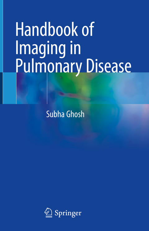
Handbook of Imaging in Pulmonary Disease
Springer International Publishing (Verlag)
978-3-030-68164-7 (ISBN)
This book is a comprehensive and easy-to-read guide to pulmonary imaging. Medical Imaging is one of the cornerstones of modern medicine, and nowhere is this more apparent than pulmonary disease. We have come a long way from the days of chest radiography, though the chest radiograph still remains the single most common imaging test ordered worldwide. Pulmonary disease is now routinely evaluated with ultra-modern computed tomography (CT), magnetic resonance imaging (MRI) and positron emission tomography (PET) scanners, while ultrasonography plays a limited role in critical care and pleural/chest wall diseases. Rapid advancements in the sub-specialty of chest imaging and an exponential increase in the knowledge of pulmonary disease have led to an increasing demand for a comprehensive yet easily digestible handbook of pulmonary imaging, which prepackages knowledge in a form that can be easily understood and readily visualized with high-quality representative images.
This book answers that need by providing the most important, relevant medical knowledge needed to handle pulmonary cases. It is divided into two sections, neoplastic disease and non-neoplastic disease. Chapters detail essential information about each disease, including presentation and the different modalities used to accurately diagnose and/or plan treatment. Major topics that are covered include bronchogenic carcinoma and other lung tumors, COPD, ILD, developmental lung disorders, pulmonary hypertension, and pulmonary infections. Each chapter includes extensive radiographic images to give a complete perspective on how these diseases present. Readers can easily see what the radiology of a particular disease entity looks like, what would be the differential diagnoses for a particular imaging abnormality, and compare the bullet review points associated with an image to their particular case.
This is an ideal guide for general and thoracic radiologists, pulmonary, sleep medicine, and critical care specialists, thoracic surgeons, as well as residents and all clinicians who treat patients with pulmonary disease.
lt;b>Subha Ghosh, MD, MBA is Clinical Assistant Professor of Radiology at Cleveland Clinic Lerner College of Medicine and Case Western Reserve University. Dr. Ghosh is a fellowship trained cardiothoracic radiologist with dual US board certifications in Internal Medicine and Radiology. He has over 10 years of experience as an academic radiologist, first as faculty at the Ohio State University, Columbus, OH from 2008- 2015 and then as Staff at the Cleveland Clinic from 2015 till date. He obtained an Executive MBA from the Weatherhead School of Management at the Case-Western Reserve University (2018-2020). He enjoys the challenge of working with complex imaging cases, teaching radiology residents and fellows, and building collaborations with interdisciplinary colleagues. He has been voted the best teacher in Thoracic Imaging at the Cleveland Clinic. Dr. Ghosh is a member of educational committees of the Radiological Society of North America and the American Board of Radiology, has published in peer-reviewed acclaimed journals and presented his work nationally and internationally. He coauthored Pulmonary Disease: Pathology, Radiology, Bronchoscopy (2020).
Section I: Neoplastic Disease.- Adenocarcinoma.- Squamous Cell Carcinoma.- Large Cell Carcinoma.- Adenosquamous Cell Carcioma.- Small cell.- Large Cell Neuroendocrine Carcinoma.- Carcinoid tumor.- Sarcomatoid carcinoma.- Salivary Gland-Type Tumors.- Papillomas.- Adenomas.- Mesenchymal Tumors.- Lymphohistiocytic Tumors.- Tumors of the Pleura.- Section II: Non-Neoplastic Disease.- Congenital Anomalies and Pediatric Lesions.- Reactive Airway Disease.- Small Airway Diseases.- Acute Lung Injury.- Common Interstitial Lung Diseases.- Other Interstitial Lung Diseases.- Pneumoconiosis.- Drug Reactions and Therapy Effects.- Vasculitis and other causes of pulmonary hemorrhage.- Pulmonary hypertension.- Viral Infections.- Bacterial Infections.- Mycobacterial Infections.- Fungal Infections.- Parasitic Infections.- Mass-like Lesions.- Lung involvement by systemic diseases.
| Erscheinungsdatum | 23.04.2021 |
|---|---|
| Zusatzinfo | XII, 235 p. 268 illus., 114 illus. in color. |
| Verlagsort | Cham |
| Sprache | englisch |
| Maße | 155 x 235 mm |
| Gewicht | 586 g |
| Themenwelt | Medizinische Fachgebiete ► Innere Medizin ► Pneumologie |
| Medizin / Pharmazie ► Medizinische Fachgebiete ► Radiologie / Bildgebende Verfahren | |
| Schlagworte | Chest Imaging • Chest Radiograph • Computed tomography • CT • MRI • PET • Pulmonary Disease • pulmonary imaging • thoracic radiology • Ultrasound |
| ISBN-10 | 3-030-68164-5 / 3030681645 |
| ISBN-13 | 978-3-030-68164-7 / 9783030681647 |
| Zustand | Neuware |
| Haben Sie eine Frage zum Produkt? |
aus dem Bereich


