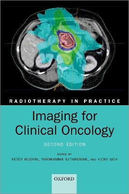
Imaging for Clinical Oncology
Oxford University Press (Verlag)
978-0-19-881850-2 (ISBN)
Imaging is a critical component in the delivery of radiotherapy to patients with malignancy, and this book teaches the principles and practice of imaging specific to radiotherapy.
Introductory chapters outline the basic principles of the available imaging modalities including x-rays, CT, ultrasound, MRI, nuclear medicine, and PET. Site specific chapters then cover the main tumour sites, reviewing optimal imaging techniques for diagnosis, staging, radiotherapy planning, and follow-up for each site. The important areas of radiation protection, exposure justification, and risks are also covered, exploring issues such as balancing radiation exposure with long-term risks of radiation effects, such as second cancer induction.
This second edition has been fully revised and updated to reflect current techniques, and includes two brand new chapters on imaging for radiotherapy treatment verification, and the role of specialist MRI techniques and functional imaging for radiotherapy planning. With insights from experts in each field and over 200 illustrations, this comprehensive and easy-to-read guide will be an invaluable resource for radiation oncologists, clinical oncologists, and radiotherapists, both qualified and in training.
ABOUT THE SERIES
Radiotherapy remains the major non-surgical treatment modality for the management of malignant disease. It is based on the application of the principles of applied physics, radiobiology, and tumour biology to clinical practice. Each volume in the series takes the reader through the basic principles of the use of ionizing radiation and then develops this by individual sites. This series of practical handbooks is aimed at physicians both training and practising in radiotherapy, as well as medical physics, dosimetrists, radiographers, and senior nurses.
Peter Hoskin has extensive clinical experience in the use of modern techniques for radiotherapy delivery. He undertakes both national and international teaching courses in the subject and is a clinical lead for the National Cancer Research Institute (NCRI) radiotherapy quality assurance programme. An experienced teacher and examiner in clinical oncology, he currently chairs the fellowship Examination Board for the Royal College of Radiologists. Peter Hoskin is the series editor for the Radiotherapy in Practice series. Thankamma Ajithkumar is a Consultant Clinical Oncologist at Addenbrooke's Hospital. He undertook a research fellowship in the Royal Marsden Hospital, London followed by speciality training in Clinical Oncology in the Eastern Deanery before becoming a Consultant Clinical Oncologist at the Bristol Haematology and Oncology Centre in 2006. In 2009 he moved to Norfolk and Norwich University Hospitals, subsequently moving to Cambridge in 2014. His research interest is to optimize the combination of radiotherapy with novel agents in paediatric brain tumours and hepato-pancreatico-biliary tumours to improve clinical outcomes. He is Chair of the European Society of Paediatric Oncology (SIOPE) Brain Tumour Radiotherapy group, and the principal investigator for the international Pediatric Normal Tissue Effects in the Clinic (PENTEC) Re-irradiation working group. He is also the radiotherapy lead for the CCLG CNS GCT subgroup and the radiotherapy research lead for the Cancer Research Network: Eastern. Vicky Goh is the Chair and Head of Department of Cancer Imaging at the School of Biomedical Engineering and Imaging Sciences, King's College London and Honorary Consultant Radiologist at Guy's and St Thomas' Hospitals, London. Her research is focused on functional imaging in cancer with CT, MRI and PET/MRI; tumour heterogeneity and AI. She has a special interest in gastrointestinal and genitourinary.
1: Peter Hoskin and Vicky Goh: Introduction
2: Connie Yip, N. Jane Taylor, Anthony Chambers, and Vicky Goh: Principles of imaging
3: Thankamma Ajithkumar, Heok Cheow, and Peter Hoskin: MRI and functional imaging in radiotherapy planning, delivery, and treatment
4: Dinos Geropantas and Victoria Ames: Breast
5: Pooja Jain, Katy Clarke, and Michael Darby: Lung and thorax
6: Peter Hoskin, Heok Cheow, and Thankamma Ajithkumar: Lymphoma
7: Kieran Foley, Carys Morgan, and Tom Crosby: Oesophageal tumours
8: Nicholas Carroll and Elizabeth C. Smyth: Gastric tumours
9: Marika Reinius, Edmund Godfrey, and Bristi Basu: Hepatic and biliary tumours
10: Thankamma Ajithkumar and David Bowden: Pancreatic tumours
11: Haesun Choi: Gastrointestinal stromal tumours
12: Vivek Misra and Rohit Kochhar: Rectal cancer
13: Rebecca Muirhead and Vicky Goh: Anal cancer
14: Ananya Choudhury and Peter Hoskin: Urological cancers
15: Kate Lankester and Lavanya Vitta: Gynaecological cancers
16: Gill Barnett and Tilak Das: Head and neck cancers
17: Sara C. Erridge, Gerard Thompson, and David Summers: Central nervous system
18: Gail Horan, Emma-Louise Gerety, Sarah Prewett, and Morag Brothwell: Soft tissue sarcomas
19: Luigi Aloj: Endocrine tumours
20: Gulshad Begum, Sarah Prewett, Gail Horan, and Emma-Louise Gerety: Primary Bone Sarcomas
21: Mark Gaze, Monique Shahid, Paul Humphries, and Francesca Peters: Paediatrics
22: Helen Addley, Katy Hickman, and Thankamma Ajithkumar: Imaging for common complications
23: June Dean: Imaging for treatment verifications
24: Simon Thomas: Radiation protection issues when imaging patients for radiotherapy
| Erscheinungsdatum | 28.09.2021 |
|---|---|
| Reihe/Serie | Radiotherapy in Practice |
| Verlagsort | Oxford |
| Sprache | englisch |
| Maße | 156 x 233 mm |
| Gewicht | 610 g |
| Themenwelt | Medizin / Pharmazie ► Gesundheitsfachberufe ► MTA - Radiologie |
| Medizin / Pharmazie ► Medizinische Fachgebiete ► Onkologie | |
| Medizinische Fachgebiete ► Radiologie / Bildgebende Verfahren ► Radiologie | |
| ISBN-10 | 0-19-881850-5 / 0198818505 |
| ISBN-13 | 978-0-19-881850-2 / 9780198818502 |
| Zustand | Neuware |
| Informationen gemäß Produktsicherheitsverordnung (GPSR) | |
| Haben Sie eine Frage zum Produkt? |
aus dem Bereich


