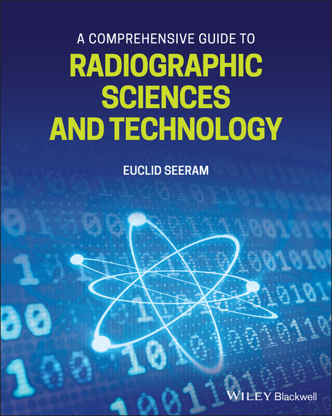
A Comprehensive Guide to Radiographic Sciences and Technology
Wiley-Blackwell (Verlag)
978-1-119-58184-0 (ISBN)
The guide begins with an overview of the radiographic sciences and technology, followed by detailed descriptions of the major components of digital radiographic imaging systems. Subsequent sections discuss the essential aspects of diagnostic radiography and computed tomography, including basic physics, imaging modalities, digital image processing, quality control, imaging informatics, and basic concepts of radiobiology and radiation protection. Throughout the book, concise chapters summarise the critical knowledge required for effective and efficient imaging of the patient while emphasising the important, yet commonly misunderstood, relationship between radiation dose and image quality.
Written by an internationally recognised expert in the field, this invaluable reference and guide:
- Provides easy access to basic physics, techniques, equipment, and safety guidelines for radiographic imaging
- Reflects the educational requirements of the American Society of Radiologic Technologists (ASRT), the Canadian Association of Medical Radiation Technologists (CAMRT), the College of Radiographers (CoR), and other radiography societies and associations worldwide
- Offers a range of pedagogical tools such as chapter outlines, key term definitions, bulleted lists, practical examples, and links to current references and additional resources
- Includes charts, diagrams, photographs, and x-ray images
»A Comprehensive Guide to Radiographic Sciences and Technology« is required reading for students in programs using ionizing radiation, those preparing for the ARRT and other global radiography certification exams, and practising technologists wanting to refresh their knowledge.
Dr Euclid Seeram, PhD, MSc, BSc, FCAMRT, currently serves as honorary senior lecturer at the University of Sydney-Faculty of Health Sciences; adjunct associate professor at Monash University-Medicine, Nursing, and Health Sciences; adjunct professor at Charles Sturt University-Faculty of Science; and Adjunct Associate Professor-Faculty of Health, University of Canberra; in Australia. He has published more than 50 papers in professional radiologic technology journals and has had 20 textbooks published on computed tomography, computers in radiology, radiographic instrumentation, digital radiography, and radiation protection. He is a founding member of the Journal of Medical Imaging and Radiation Sciences and is now on editorial boards for Radiography; Biomedical Imaging and Intervention Journal; Open Journal of Radiology; Journal of Allied Health; Journal of Social Science & Allied Health Professions. Euclid also serves on the international advisory panel for the Journal of Medical Radiation Sciences.
Foreword
Preface
SECTION 1: INTRODUCTION
Chapter 1: Radiographic Sciences and Technology: An Overview
Radiographic Imaging Systems: Major Modalities and Components
Radiographic Imaging Physics
Essential Physics of Diagnostic Imaging
Digital Radiographic Imaging Modalities
Radiographic Exposure Technique
Image Quality Considerations
Computed Tomography-Physics and Instrumentation
Quality Control
Imaging Informatics at a Glance
Radiation Protection and Dose Optimization
Radiobiology
Technical Factors Affecting Dose in Radiographic Imaging
Radiation Protection in Diagnostic Radiography
Radiation Protection Regulations
Optimization of Radiation Protection
References
Chapter 2: Digital Radiographic Imaging Systems: Major Components
Film-Screen Radiography: A Short Review of Principles
Digital Radiography Modalities: Major System Components
Computed Radiography
Flat-Panel Digital Radiography
Digital Fluoroscopy
Digital Mammography
Computed Tomography
Image Communication Systems
Picture Archiving and Communication System
References
SECTION 2: BASIC RADIOGRAPHIC SCIENCES and TECHNOLOGY
Chapter 3: Basic Physics of Diagnostic Radiography
Structure of the Atom
Nucleus
Electrons, Quantum Levels, Binding Energy, Electron Volts
Energy Dissipation in Matter
Excitation
Ionization
Types of Radiation
Electromagnetic
Particulate
X-Ray Generation
X-Ray Production
Properties of X-rays
Origin of X-Rays
Characteristic Radiation
Bremstrahlung Radiation
X-ray Emission
X-Ray Beam Quantity and Quality
Factors Affecting X-Ray Beam Quantity and Quality
Interaction of Radiation with Matter
Mechanisms of Interaction in Diagnostic X-Ray Imaging
Radiation Attenuation
Linear Attenuation Coefficient
Mass Attenuation Coefficient
Half Value Layer
Radiation Quantities and Units
References
Chapter 4: The X-Ray Tube and Generator
Physical Components of the X-Ray Machine
Components of the X-Ray Circuit
The Power Supply to the X-Ray Circuit
The Low Voltage Section (Control Console)
The High Voltage Section
Types of X-Ray Generators
Three-Phase Generators
High-Frequency Generators
Power Ratings
The X-Ray Tube: Structure and Function
Major Components
Special X-Ray Tubes: Basic Design Features
Double-Bearing Axle
Heat Capacity and Heat Dissipation Considerations
X-Ray Beam Filtration and Collimation
Inherent and Added Filtration
Effects of Filtration on X-ray Tube Output Intensity
Half-Value Layer
Collimation
References
Chapter 5: Digital Image Processing at a Glance
Digital Image Processing
Definition
Image Formation and Representation
Processing Operations
Characteristics of Digital Images
Gray Scale Processing
Windowing
Conclusion
References
Chapter 6: Digital Radiographic Imaging Modalities: Principles and Technology
Computer Radiography
Essential Steps
Basic Physical Principles
Response of the IP to Radiation Exposure
Standardized Exposure Indicator
Flat-Panel Digital Radiography
What is Flat-Panel Digital Radiography (FPDR)?
Types of FPDR Systems
Basic Physical Principles of Indirect and Direct Flat-Panel Detectors
The Fill Factor of the Pixel in the Flat-Panel Detector
Exposure Indicator
Image Quality Descriptors for DR Systems
Continuous Quality Improvement for DR Systems
Digital Fluoroscopy
Digital Fluoroscopy Modes
Image Intensifier-Based Digital Fluoroscopy Characteristics
Flat-Panel Digital Fluoroscopy Characteristics
Digital Mammography
Screen-Film Mammography: Basic Principles
Full-Field Digital Mammography-Major Elements
Digital Tomosynthesis at a Glance
Imaging System Characteristics
Synthesized 2D Digital Mammography
References
Chapter 7: Image Quality and Dose
The process of Creating an Image
Image Quality Metrics
Contrast
Contrast Resolution
Spatial Resolution
Noise
Contrast-to-Noise Ratio
Signal-to-Noise Ratio
Artifacts
Dose and Image Quality
Digital Detector Response to the Dose
Detective Quantum Efficiency
References
SECTION 3: COMPUTED TOMOGRAPHY: BASIC PHYSICS and TECHNOLOGY
Chapter 8: The Essential Technical Aspects of Computed Tomography
Physics
Radiation Attenuation
Technology
Data Acquisition: Principles and Components
Image Reconstruction
Image Display, Storage, and Communication
MultiSlice CT (MSCT): Principles and Technology
Slip-Ring Technology
X-Ray Tube Technology
Interpolation Algorithms
MSCT Detector Technology
Selectable Scan Parameters
Isotropic CT Imaging
MSCT Image Processing
Image Postprocessing
Windowing
3-D Image Display Techniques
Image Quality
Spatial Resolution
Contrast Resolution
Noise
Radiation Protection
CT Dosimetry
Factors Affecting Patient Dose
Optimizing Radiation Protection
Conclusion
References
SECTION 4: CONTINUOUS QUALITY IMPROVEMENT
Chapter 9: Fundamentals of Quality Control
Introduction
Definitions
Essential Steps of QC
QC Responsibilities
Steps in Conducting a QC Test
The Tolerance Limits or Acceptance Criteria
Parameters for QC Monitoring
QC Testing Frequency
Tools for QC Testing
The Format of a QC Test
Performance Criteria/Tolerance Limits for Common QC Tests
Radiography
Fluoroscopy
Repeat Image Analysis
Computed Tomography QC Tests for Technologists
References
SECTION 5: PACS and IMAGING INFORMATICS
Chapter 10: Imaging Informatics at a Glance
Introduction
Picture Archiving and Communication Systems: Characteristic Features
Definition
Core Technical Components
Imaging Informatics?
Enterprise Imaging
Cloud Computing
Big Data
Artificial Intelligence
Machine Learning and Deep Learning
Applications in Medical Imaging
AI in CT Image Reconstruction
Ethics of AI in Radiology-A Joint Multi-Society Summary Statement.
References
SECTION 5: RADIATION PROTECTION
Chapter 11: Basic Concepts of Radiobiology
What is Radiobiology?
Generalizations About Radiation Effects on Living Organisms
Relevant Physical Processes
Ionization
Excitation
Linear Energy Transfer
Relative Biological Effectiveness
Radiolysis of Water
Dose-Response Models
The Linear Non-Threshold (LNT) Dose-Response Model
The Linear Threshold Dose-Response Model
Stochastic Effects
Deterministic Effects
Radiation Effects on the Conceptus
References
Chapter 12: Technical Dose Factors in Radiography, Fluoroscopy, and CT
Dose Factors in Digital Radiography
The X-Ray Generator
Exposure Technique Factors
X-Ray Beam Filtration
Collimation and Field Size
SID and SSD
Patient Thickness and Density
Scattered Radiation Grid
The Sensitivity of the Image Receptor
Dose Factors in Fluoroscopy
Fluoroscopic Exposure Factors
Fluoroscopic Equipment Factors
CT Radiation Dose Factors and Dose Optimization Considerations
Dose Distribution in the Patient
CT dose Metrics
Factors Affecting the Dose in CT
Dose Optimization Overview
References
Chapter 13: Essential Principles of Radiation Protection
Introduction
Why Radiation Protection?
Categories of Data from Human Exposure
Radiation Dose-Risk Models
Summary of Biological Effects
Radiation Protection Organizations
Objectives of Radiation Protection
Radiation Protection Philosophy
ICRP Radiation Protection Framework
Justification
Optimization
Dose Limits
Dose Limits
Personal Actions
Time
Shielding
Distance
Radiation Quantities and Units
Sources of Radiation Exposure
Quantities and Units
Personnel Dosimetry
Optimization of Radiation Protection
Regulatory and Guidance Recommendations
Diagnostic Reference Levels
Gonadal Shielding: Past Considerations
X-Ray Protective Shielding
Current State of Gonadal Shielding
References
Index
| Erscheinungsdatum | 11.06.2021 |
|---|---|
| Verlagsort | Hoboken |
| Sprache | englisch |
| Maße | 203 x 252 mm |
| Gewicht | 588 g |
| Einbandart | kartoniert |
| Themenwelt | Medizin / Pharmazie ► Allgemeines / Lexika |
| Medizin / Pharmazie ► Gesundheitsfachberufe ► MTA - Radiologie | |
| Medizinische Fachgebiete ► Radiologie / Bildgebende Verfahren ► Kernspintomographie (MRT) | |
| Medizinische Fachgebiete ► Radiologie / Bildgebende Verfahren ► Radiologie | |
| ISBN-10 | 1-119-58184-2 / 1119581842 |
| ISBN-13 | 978-1-119-58184-0 / 9781119581840 |
| Zustand | Neuware |
| Informationen gemäß Produktsicherheitsverordnung (GPSR) | |
| Haben Sie eine Frage zum Produkt? |
aus dem Bereich


