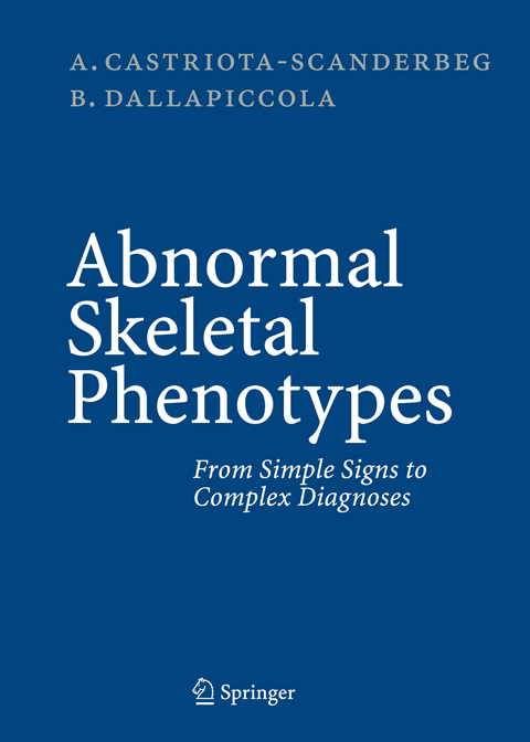
Abnormal Skeletal Phenotypes
Springer Berlin (Verlag)
978-3-540-67997-4 (ISBN)
I.- Skull.- Thorax.- Spine.- Pelvis.- Long Bones.- Hands.- Feet.- Joints.- Generalized Skeletal Abnormalities.- II.- Aarskog Syndrome.- Achondrogenesis, Type IB.- Achondrogenesis, Type II.- Achondroplasia.- Acrofacial Dysostosis, Nager Type.- Angelman Syndrome.- Apert Syndrome.- Asphyxiating Thoracic Dysplasia.- Atelosteogenesis.- Bardet-Biedl Syndrome.- Beckwith-Wiedemann Syndrome.- C Syndrome.- Campomelic Dysplasia.- Carpenter Syndrome.- Cerebro-costo-mandibular Syndrome.- CHARGE Association.- Chondrodysplasia Punctata, Conradi-Hünermann Type.- Chondrodysplasia Punctata, Rhizomelic Type.- Chondrodysplasia Punctata, Brachytelephalangic Type.- Chondroectodermal Dysplasia.- Chromosome 4p- Syndrome.- Chromosome Trisomy 13 Syndrome.- Chromosome Trisomy 18 Syndrome.- Chromosome Trisomy 21 Syndrome.- Cleidocranial Dysplasia.- Cockayne Syndrome.- Coffin-Lowry Syndrome.- Coffin-Siris Syndrome.- Cohen Syndrome.- Craniometaphyseal Dysplasia, Dominant Type.- Cri-du-chat Syndrome.- Crouzon Syndrome.- De Lange Syndrome.- Diaphyseal Dysplasia.- Diastrophic Dysplasia.- Dubowitz Syndrome.- Dyschondrosteosis.- Dysosteosclerosis.- Ectodermal Dysplasias.- Ehlers-Danlos Syndromes.- Enchondromatosis.- Exostoses, Multiple.- Fanconi Anemia.- Focal Dermal Hypoplasia Syndrome.- Freeman-Sheldon Syndrome.- Frontometaphyseal Dysplasia.- Goldenhar Syndrome.- Hallermann-Streiff Syndrome.- Holt-Oram Syndrome.- Kenny-Caffey Syndrome.- Klippel-Feil Anomaly.- Klippel-Trenaunay-Weber Syndrome.- Kniest Dysplasia.- Larsen Syndrome.- Marfan Syndrome.- McCune-Albright Syndrome.- Meckel Syndrome.- Melnick-Needles Syndrome.- Melorheostosis.- Mental Retardation, X-Linked, Associated with FRA Xq27.3.- Mesomelic Dwarfism, Langer Type.- Mesomelic Dwarfism, Nievergelt Type.- Metatropic Dysplasia.- MultipleEpiphyseal Dysplasia.- Nail-Patella Syndrome.- Nevoid Basal Cell Carcinoma Syndrome.- Noonan Syndrome.- Opitz Syndrome.- Oro-facio-digital Syndrome, Type I.- Oro-facio-digital Syndrome, Type II.- Osteogenesis Imperfecta, Type I.- Osteogenesis Imperfecta, Type IIA.- Osteogenesis Imperfecta, Type IIB/III.- Osteopathia Striata with Cranial Sclerosis.- Osteopetrosis, Infantile Type.- Osteopetrosis, Adult Type.- Osteopoikilosis.- Oto-palato-digital Syndrome, Type I.- Oto-palato-digital Syndrome, Type II.- Pena-Shokeir Syndrome.- Pfeiffer Syndrome.- Poland Syndrome.- Prader-Willi Syndrome.- Progeria.- Pseudoachondroplasia.- Pyknodysostosis.- Roberts Syndrome.- Robin Sequence.- Robinow Syndrome.- Rubinstein-Taybi Syndrome.- Saethre-Chotzen Syndrome.- Seckel Syndrome.- Short Rib-Polydactyly Syndrome, Type I.- Short Rib-Polydactyly Syndrome, Type II.- Silver-Russell Syndrome.- Smith-Lemli-Opitz Syndrome.- Sotos Syndrome.- Spondyloepimetaphyseal Dysplasia, Irapa Type.- Spondyloepimetaphyseal Dysplasia, Strudwick Type.- Spondyloepiphyseal Dysplasia Congenita.- Spondyloepiphyseal Dysplasia Tarda.- Spondylometaphyseal Dysplasia, Kozlowski Type.- Stickler Syndrome.- Thanatophoric Dysplasia.- Thrombocytopenia-Absent Radius Syndrome.- Treacher-Collins Syndrome.- Tricho-rhino-phalangeal Syndrome, Type I.- Tricho-rhino-phalangeal Syndrome, Type II.- Turner Syndrome.- VATER Association.- Williams Syndrome.
From the reviews:
5 Stars Doody's review:
Description: This is a new and exciting book that provides a review of skeletal abnormalities in two ways. The authors approach each abnormal radiologic finding and link each abnormality to specific genetic disorders in the first section. In the second section, the authors review 111 common skeletal dysmorphologic conciliations. Each entry is followed by excellent radiologic images.
Audience: ... this is a great teaching tool for pediatrics, radiology, and genetics residents as well for medical students in general. This is a "must have" book for the pediatric radiologist in practice.
Assessment: The authors have accomplished a tremendous task. They present us with a unique book that deviates from the traditional radiologic atlas format. It allows clinicians to extract from radiologic reports findings that may be significant and associated with specific genetic conditions. This is a unique book, not only in its format, but also in the information it presents. It is the basis of radiological skeletal dysmorphology. The printing, presentation, and images presented are of excellent quality.
"This is a concise book, which can be used as a reference for trying to analyse skeletal dysplasias. ... There are many good quality clinical pictures and radiographs. ... The text is thoroughly referenced. The text would be useful for orthopaedic, paediatric and radiology departments and all health care professionals, especially orthopaedics and radiologists." (Dr. P. Richards, RAD Magazine, May, 2006)
"This is an excellent book, written by two experienced, well known paediatric radiologists. The book is divided into two parts. ... The images are of high quality and are relevant to the text. I highly recommend this book to all hospitals and large private practice libraries. It is an indispensable book for those working in paediatric radiology, orthopaedic surgery, paediatric and genetics."(K Kozlowski, Australasian Radiology, Vol. 51, 2007)
| Erscheint lt. Verlag | 26.10.2005 |
|---|---|
| Zusatzinfo | XIV, 962 p. |
| Verlagsort | Berlin |
| Sprache | englisch |
| Maße | 193 x 260 mm |
| Gewicht | 2840 g |
| Themenwelt | Medizinische Fachgebiete ► Radiologie / Bildgebende Verfahren ► Radiologie |
| Schlagworte | Birth Defects • Chromosom • Diagnosis • Genetics • Malformation Syndromes • Osteogenesis imperfecta • Radiology • Skeletal Dysplasias • Skelett |
| ISBN-10 | 3-540-67997-9 / 3540679979 |
| ISBN-13 | 978-3-540-67997-4 / 9783540679974 |
| Zustand | Neuware |
| Informationen gemäß Produktsicherheitsverordnung (GPSR) | |
| Haben Sie eine Frage zum Produkt? |
aus dem Bereich


