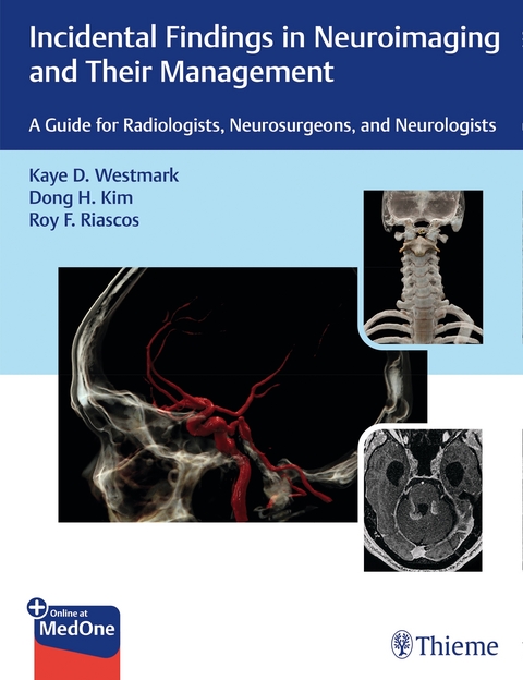
Incidental Findings in Neuroimaging and Their Management
Thieme (Verlag)
978-1-62623-828-2 (ISBN)
»Incidental Findings in Neuroimaging and Their Management: A Guide for Radiologists, Neurosurgeons, and Neurologists« presents a streamlined, case-based approach to 50 commonly seen incidental findings in neuroimaging. Edited by Kaye Westmark, Dong Kim, and Roy Riascos, this unique book provides the necessary knowledge to manage significant unexpected findings—from identification and analysis to efficacious interventions. With collaborative contributions from neuroradiologists, neurosurgeons, neurologists, otolaryngologists, body and musculoskeletal imaging experts, endocrinologists and hematologists/oncologists, this resource encompasses a wide spectrum of incidental findings.
Organized by six sections, the book starts with normal variants that are extremely important to recognize in order to avoid unwarranted additional testing and unnecessary stress for the patient. Subsequent sections detail abnormalities that require extensive clinical evaluation in order to determine ideal management. These include incidental findings for extracranial, extra-spinal, intracranial, and intraspinal imaging. The final section outlines CT and MR imaging artifacts that are particularly concerning because they may mimic more dangerous pathologies while degrading imaging quality and obscuring real findings.
Key Features
- Key findings and differential diagnosis are listed for each entity
- Diagnostic decision trees present algorithms in an easy-to-understand manner
- Artifact analyses explain the technical reason for each artifact and what can be done to mitigate effects
- Clinical Q&As connect the radiologic diagnosis with actual case management decisions and provide in-depth background information that is applicable to management in various scenarios
This essential guide will help trainee and practicing neuroradiologists, neurosurgeons, and neurologists interpret incidental spine and brain imaging findings and make clinically informed, complex treatment decisions.
This book includes complimentary access to a digital copy on https://medone.thieme.com.
Section I: Normal Variants
Chapter 1 Arachnoid Granulation
Chapter 2 Asymmetrical Lateral Ventricles
Chapter 3 Basal Ganglia/Dentate Nuclei Mineralization
Chapter 4 Cavum Septum Pellucidum
Chapter 5 Choroid Fissure Cyst
Chapter 6 Empty Sella Configuration
Chapter 7 High-Riding Jugular Bulb
Chapter 8 Hyperostosis Frontalis Interna
Chapter 9 Prominent Perivascular Space
Chapter 10 Simple Pineal Cyst
Section II: Intracranial Incidental Findings
Chapter 11 Diffuse White Matter Hyperintensities
Chapter 12 Brain Capillary Telangiectasias
Chapter 13 Developmental Venous Anomaly
Chapter 14 Cerebral Cavernous Malformations
Chapter 15 Colloid Cyst
Chapter 16 Arachnoid Cysts
Chapter 17 Mega Cisterna Magna
Chapter 18 Benign Enlargement of Subarachnoid Spaces
Chapter 19 Pituitary Incidentaloma and Incidental Silent Macroadenoma
Chapter 20 Pituitary Gland: Diffuse Enlargement
Chapter 21 Incidental Glial Neoplasms
Chapter 22 Incidental Meningioma
Section III: Head and Neck Related Incidental Findings
Chapter 23 Head and Neck-Related Incidental Findings
Section IV: Spinal Incidental Findings
Chapter 24 Os Odontoideum
Chapter 25 Tarlov's Cyst
Chapter 26 Approach to the Solitary Vertebral Lesion on Magnetic Resonance Imaging
Chapter 27 Diffusely Abnormal Marrow Signal within the Vertebrae on MRI
Chapter 28 Filum Terminale Lipoma
Chapter 29 Ventriculus Terminalis
Chapter 30 Prominent Central Canal
Chapter 31 Low-Lying Conus
Chapter 32 Incidental Solitary Sclerotic Bone Lesion
Section V: Extraspinal Incidental Findings
Chapter 33 Abdominal Aortic Aneurysm
Chapter 34 Renal Mass
Chapter 35 Renal Cyst
Chapter 36 Thyroid Mass
Chapter 37 Adrenal Mass
Chapter 38 Retroperitoneal Lymph Nodes
Chapter 39 Incidental Pelvic Mass
Section VI: Artifacts That May Simulate Disease Entities and Obscure Pathology
Chapter 40 Computed Tomography Artifacts
Chapter 41 MRI Patient-Related Motion Artifacts
Chapter 42 Magnetic Susceptibility-Related Artifacts on MRI
Chapter 43 MRI Technical and Sequence-Specific Artifacts
| Erscheinungsdatum | 04.08.2020 |
|---|---|
| Zusatzinfo | 683 Illustrations |
| Verlagsort | Stuttgart |
| Sprache | englisch |
| Maße | 216 x 280 mm |
| Gewicht | 1361 g |
| Einbandart | gebunden |
| Themenwelt | Medizinische Fachgebiete ► Chirurgie ► Neurochirurgie |
| Medizin / Pharmazie ► Medizinische Fachgebiete ► Neurologie | |
| Medizinische Fachgebiete ► Radiologie / Bildgebende Verfahren ► Neuroradiologie | |
| Medizinische Fachgebiete ► Radiologie / Bildgebende Verfahren ► Nuklearmedizin | |
| Medizinische Fachgebiete ► Radiologie / Bildgebende Verfahren ► Radiologie | |
| Schlagworte | brain imaging • neuroimaging • neurosurgery • radiologic diagnosis • spine imaging |
| ISBN-10 | 1-62623-828-6 / 1626238286 |
| ISBN-13 | 978-1-62623-828-2 / 9781626238282 |
| Zustand | Neuware |
| Informationen gemäß Produktsicherheitsverordnung (GPSR) | |
| Haben Sie eine Frage zum Produkt? |
aus dem Bereich


