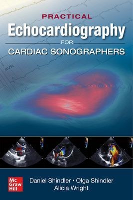
Practical Echocardiography for Cardiac Sonographers
McGraw-Hill Education (Verlag)
978-1-260-45779-7 (ISBN)
A practical, visual, authoritative quick-reference guide to mastering echocardiography
The most efficient and thorough way to learn echocardiography is visually. By seeing and comparing different variations of important echo findings, you shorten the learning process and build retention at the same time. Covering the essential techniques, standards, and cardiac disorders you need to know, Practical Echocardiography for Cardiac Sonographers is an innovative guide that takes you step by step through the scanning process in different disorders, helps you accurately interpret and clinically apply echocardiographic information, describes the physics of ultrasound in easy-to-understand language, and covers common and rare relevant cardiac conditions.
Formatted in a way that makes finding the right answers quick and easy, Echocardiography for Cardiac Sonographers delivers comprehensive fully rounded, interactive education that will get you up to speed on this critically important discipline in no time.
This comprehensive guide covers:
Heart FailureCoronary Artery DiseaseAortic Valve DiseaseMitral valve DiseaseProsthetic ValvesHypertrophic CardiomyopathyPericardial DiseaseEndocarditisCardiac Tumors and MassesPulmonary DisordersTricuspid and Pulmonic ValvesAorta and Congenital Heart DisordersStroke
Daniel Shindler, MD FACC is Professor of Medicine, Rutgers-Robert Wood Johnson Medical School, New Brunswick, NJ, and Governor, New Jersey Chapter American College of Cardiology. Olga Shindler, MD is President, Mobile Cardiac Ultrasound, East Brunswick, NJ Alicia Wright, RDCS is a cardiac sonographer at Robert Wood Johnson University Hospital.
Table of Contents
Introduction
1. Physics of ultrasound
2. Imaging cardiac anatomy
3. Principles of Doppler
4. Echocardiographic measurements
5. Echocardiographic calculations
6. Echocardiographic instrument settings
7. Contrast ultrasound
8. Bubble physics and instrument settings
9. Tissue Doppler
10. Myocardial strain
11. Point of care ultrasound
12. Transesophageal imaging
13. Intracardiac and transcranial ultrasound
14. Vascular ultrasound
15. Left ventricular function
16. Diastology
17. Coronary artery disease
18. Wall motion abnormalities
19. Exercise stress testing
20. Pharmacologic stress testing
21. Myocardial perfusion
22. Complications of myocardial infarction
23. Systemic hypertension
24. Pulmonary disorders
25. Pulmonary hypertension
26. Evaluation of dyspnea
27. Valvular heart disease
28. Aortic stenosis
29. Aortic regurgitation
30. Mitral regurgitation
31. Mitral and tricuspid stenosis
32. Tricuspid regurgitation
33. Pulmonic valve regurgitation
34. Pulmonic valve stenosis
35. Heart failure
36. Cardiomyopathies
37. Dilated cardiomyopathy
38. Hypertrophic cardiomyopathy
39. Infiltrative cardiomyopathy
40. Disorders of the aorta
41. Aortic aneurysm
42. Aortic dissection
43. Endocarditis
44. Pericardial diseases
45. Pericardial effusion and tamponade
46. Pericardial constriction
47. Preoperative clearance
48. Atrial fibrillation and flutter
49. Imaging in structural heart disease
50. Cardiac tumors and masses
51. Cardio oncology
52. Imaging in the stroke patient
53. Imaging in rheumatological disorders
54. Imaging in the intensive care unit
55. Imaging in the emergency room
56. Imaging the elderly patient
57. Pregnancy
58. Pediatric imaging
59. Congenital heart disease
60. Atrial septal defects
61. Ventricular septal defects
62. Transposition of the great vessels
63. Congenital disorders of the tricuspid valve
64. Nomenclature in congenital heart disorders
65. Postoperative congenital heart disease
66. The stethoscope in echocardiography
67. The electrocardiogram in echocardiography
68. The chest x-ray
69. CT scan of the chest
70. MRI imaging of the heart
71. Internet resources
72. Guidelines
References
| Erscheinungsdatum | 09.07.2020 |
|---|---|
| Zusatzinfo | 60 Illustrations |
| Verlagsort | OH |
| Sprache | englisch |
| Maße | 152 x 226 mm |
| Gewicht | 531 g |
| Themenwelt | Medizin / Pharmazie ► Allgemeines / Lexika |
| Medizinische Fachgebiete ► Innere Medizin ► Kardiologie / Angiologie | |
| ISBN-10 | 1-260-45779-6 / 1260457796 |
| ISBN-13 | 978-1-260-45779-7 / 9781260457797 |
| Zustand | Neuware |
| Haben Sie eine Frage zum Produkt? |
aus dem Bereich


