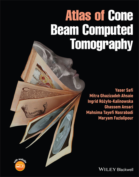
Atlas of Cone Beam Computed Tomography
Wiley-Blackwell (Verlag)
978-1-119-66777-3 (ISBN)
»Atlas of Cone Beam Computed Tomography« delivers a robust collection of cases using this advanced method of imaging for oral and maxillofacial radiology. The book features over 1,500 high-quality CBCT scans with succinct descriptions covering a wide range of maxillofacial region conditions, including normal anatomy, anomalies, inflammatory diseases, and degenerative diseases.
Easy to navigate and featuring multiple images of normal variation and pathologies, the book offers readers guidance on the diagnostic values of CBCT, as well as CBCT images of the inferior alveolar nerve canal, dental implants, temporomandibular joint evaluations, and surgical interventions. The book also includes:
- A thorough introduction to cone beam computed tomography, including in vivo and in vitro preparation and evaluation, indications in dentistry, and indications in medicine
- Comprehensive explorations of cone beam computed tomography artefacts and anatomic landmarks
- Practical discussions of cone beam computed tomography of dental structure, including normal anatomy, anomalies, and the difficulties of eruption
- In-depth examinations of cone beam computed tomography of pathological growth and development, including maxillofacial congenital and developmental anomalies
Perfect for graduate dental students and postgraduate dental students in oral and maxillofacial radiology, »Atlas of Cone Beam Computed Tomography« is also useful to general dentists, oral and maxillofacial radiologists, head and neck maxillofacial surgeons, head and neck radiologists, general radiologists, and ENT surgeons.
Yaser Safi, DDS, MSc, Associate Professor, Department of Oral and Maxillofacial Radiology, School of Dentistry, Shahid Beheshti University of Medical Sciences, Tehran, Iran.
Mitra Ghazizadeh Ahsaie, DDS, MSc, MPH, Assistant Professor, Department of Oral and Maxillofacial Radiology, School of Dentistry, Shahid Beheshti University of Medical Sciences, Tehran, Iran.
Ingrid Rozylo-Kalinowska, MD, PhD, President of the European Academy of Dentomaxillofacial Radiology, Regional Director of the International Association of Dento-Maxillo-Facial Radiology, Vice-President of the Polish Dental Association, Department of Dental and Maxillofacial Radiodiagnostics, Faculty of Medical Dentistry, Medical University of Lublin, Lublin, Poland.
Ghassem Ansari, DDS, MSc, PhD (Glasgow), FHD (UCLA), Professor, Head of the Hospital Dentistry and Sedation Unit, Department of Pediatric Dentistry, School of Dentistry, Shahid Beheshti University of Medical Sciences, Tehran, Iran.
Mahsima Tayefi Nasrabadi, DDS, MSc, Assistant Professor, Department of Oral and Maxillofacial Radiology, Dental College, Alborz University of Medical Sciences, Karaj, Iran.
Maryam Fazlalipour, DDS, MSc, Assistant Professor, Department of Oral and Maxillofacial Radiology, School of Dentistry, Tehran University of Medical Sciences, Tehran, Iran.
Preface
About the Companion Website
1. CBCT Introduction
1-1 Science, Preparation & Evaluations
1-2 Indications in Dentistry
1-3 Indications in Medicine
2. CBCT and Artifacts
3. Anatomic Landmarks
3-1 Normal
3-2 variations
4. CBCT of Dental Structure
4-1 Normal Anatomy & anomalies
4-2 Difficulties of Eruption
5. CBCT of Congenital and Developmental maxillofacial anomalies
6. CBCT of Maxillofacial Trauma
6-1 Dental Fracture
6-2 Dento-Alveolar Fracture
6-3 Bone Fractures
7. CBCT and Soft Tissue Calcifications and Ossifications
8. CBCT of Foreign Bodies
9. CBCT and its Diagnostic Values in:
9-1 Endodontics
9-2 Periodontics
9-3 Orthodontics
10. CBCT and Maxillofacial Pathology Assessment:
10-1 Odontogenic Lesions
10-2 Non-odontogenic Lesions
11.CBCT and ENT Assessment
12. CBCT and Inferior Alveolar Nerve Canal
13. CBCT of Dental Implants
13-1 Pre-surgical Implant Assessments
13-2 Post operative Complications
14. CBCT and TMJ Evaluations
14-1 Initial Evaluations
14-2 Post treatment evaluation
15. Interventional CBCT
Conclusion
Bibliography and Further Readings
| Erscheinungsdatum | 07.03.2022 |
|---|---|
| Verlagsort | Hoboken |
| Sprache | englisch |
| Maße | 223 x 277 mm |
| Gewicht | 1514 g |
| Einbandart | gebunden |
| Themenwelt | Medizin / Pharmazie ► Medizinische Fachgebiete |
| Medizin / Pharmazie ► Zahnmedizin ► Klinik und Praxis | |
| Schlagworte | Cone-Beam Computed Tomography • cone beam CT • zahnärtzliche Radiologie |
| ISBN-10 | 1-119-66777-1 / 1119667771 |
| ISBN-13 | 978-1-119-66777-3 / 9781119667773 |
| Zustand | Neuware |
| Haben Sie eine Frage zum Produkt? |
aus dem Bereich


