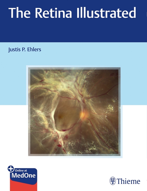
The Retina Illustrated
Thieme Medical Publishers Inc (Verlag)
978-1-62623-831-2 (ISBN)
The diagnosis and treatment of retinal eye disorders often presents significant clinical challenges. »The Retina Illustrated«, edited by renowned retina specialist Justis Ehlers and an impressive group of worldwide contributors, provides a rapid-fire yet thorough approach to the visual world of retinal disease. Organized into ten sections and 102 succinct yet comprehensive chapters, this richly illustrated reference covers the full spectrum of retinal disorders, ranging from common degenerative diseases to emerging infectious retinal diseases.
The book opens with a discussion of state-of-the-art diagnostic tools, followed by nine disorderspecific sections describing diagnosis and treatment of a wide-spectrum of retinal disorders, including degenerative, vascular, infectious, inflammatory, traumatic, oncology, and toxicities. The text covers the full age continuum, from conditions primarily impacting older adults, such as age-related macular degeneration and choroidal atrophy, to pediatric disorders, such as retinopathy of prematurity.
Key Features:
- Discussion of cutting-edge imaging diagnostics, including ultra-widefield angiography, intraoperative optical coherence tomography, and OCT angiography
- More than 400 high-quality illustrations augment the text, enhancing understanding of retinal disease, from symptoms and signs to differential diagnosis and management
- Reader-friendly format provides rapid assessment and review of numerous conditions
This is a must-have reference for all providers who encounter patients with retinal disease, including general ophthalmologists, retina specialists, emergency medicine physicians, and optometrists. Ophthalmology residents and fellows-in-training will also find this book an invaluable education tool.
This book includes complimentary access to a digital copy on https://medone.thieme.com.
Part I Retinal Diagnostics
1 Optical Coherence Tomography
2 Fluorescein Angiography
3 Indocyanine Green Angiography
4 Optical Coherence Tomography Angiography
5 Intraoperative Optical Coherence Tomography
6 Fundus Autofluorescence
Part II Macular and Peripheral Vitreoretinal Interface Disorders
7 Posterior Vitreous Detachment
8 Epiretinal Membrane
9 Macular Holes
10 Vitreomacular Traction Syndrome
11 Retinal Tears and Holes
12 Primary Rhegmatogenous Retinal Detachment
13 Giant Retinal Tear
14 Retinal Dialysis
15 Proliferative Vitreoretinopathy
16 Retinoschisis
17 Lattice Degeneration
Part III Retinal Vascular Disease
18 Diabetic Retinopathy and Diabetic Macular Edema
19 Retinal Vein Occlusion
20 Retinal Artery Occlusion
21 Ocular Ischemic Syndrome
22 Retinal Arterial Macroaneurysm
23 Radiation Retinopathy
24 Sickle Cell Retinopathy
25 Hypertensive Retinopathy
Part IV Retinal Degenerations and Dystrophies
26 Dry Age-Related Macular Degeneration
27 Wet Age-Related Macular Degeneration
28 Polypoidal Choroidal Vasculopathy
29 Age-Related Choroidal Atrophy
30 Myopic Degeneration and Myopic Foveoschisis
31 Angioid Streaks
32 Best Disease
33 Adult Vitelliform Macular Dystrophy
34 Pattern Dystrophy
35 Macular Telangiectasia
36 Retinitis Pigmentosa
37 Cone Dystrophy
38 Paraneoplastic Retinopathies
39 Congenital Stationary Night Blindness
40 Albinism
41 Gyrate Atrophy
42 Choroideremia
43 Stargardt Disease
44 Cobblestone Degeneration
Part V Other Macular Disorders
45 Central Serous Chorioretinopathy and Pachychoroid Spectrum
46 Hypotony Maculopathy
47 Cystoid Macular Edema
48 Choroidal Folds
49 Optic Pit-Related Maculopathy
Part VI Chorioretinal Infectious and Inflammatory Disorders
50 Bacterial Endophthalmitis
51 Fungal Endophthalmitis
52 Toxoplasmosis
53 Toxocariasis
54 Acute Retinal Necrosis
55 Syphilitic Uveitis
56 Ocular Tuberculosis
57 Cytomegalovirus Retinitis
58 Human Immunodeficiency Virus Retinopathy
59 West Nile Retinopathy
60 Ebola Virus
61 Zika Virus and the Retina
62 Presumed Ocular Histoplasmosis Syndrome
63 Diffuse Unilateral Subacute Neuroretinitis
64 Multifocal Choroiditis and Panuveitis
65 Ocular Sarcoidosis
66 Serpiginous Choroiditis
67 Birdshot Retinochoroiditis
68 Multiple Evanescent White Dot Syndrome
69 Acute Posterior Multifocal Placoid Pigment Epitheliopathy
70 Behcet Disease
71 Pars Planitis
72 Vogt-Koyanagi-Harada Syndrome
73 Sympathetic Ophthalmia
74 Susac Syndrome
Part VII Retinal Trauma and Other Conditions
75 Traumatic Macular Hole
76 Commotio Retinae
77 Choroidal Rupture
78 Chorioretinitis Sclopetaria
79 Dislocated Lens
80 Intraocular Foreign Body
81 Terson Syndrome
82 Purtscher and Purtscher-like Retinopathy
83 Laser Maculopathy
84 Solar Retinopathy
85 Choroidal Detachments
Part VIII Toxic and Drug-Related Retinopathies
86 Hydroxychloroquine Retinopathy
87 Ocriplasmin Retinopathy
88 Hemorrhagic Occlusive Retinal Vasculitis Secondary to Drug Exposure
89 Talc Retinopathy
90 Tamoxifen Retinopathy
91 Retinopathy Secondary to Targeted Cancer Therapies
Part IX Posterior Segment Tumors
92 Choroidal Nevus
93 Choroidal Melanoma
94 Choroidal Metastasis
95 Intraocular Lymphoma
96 Retinal Vascular Tumors
97 Retinoblastoma
98 Hamartomas of the Retina: Astrocytic and Retina/Retinal Pigment Epithelium
Part X Pediatric Vitreoretinal Disease
99 Coats Disease
100 Retinopathy of Prematurity
101 Familial Exudative Vitreoretinopathy
102 Persistent Fetal Vasculature
| Erscheinungsdatum | 16.11.2019 |
|---|---|
| Zusatzinfo | 407 Abbildungen |
| Verlagsort | New York |
| Sprache | englisch |
| Maße | 216 x 277 mm |
| Gewicht | 1451 g |
| Einbandart | kartoniert |
| Themenwelt | Medizin / Pharmazie ► Medizinische Fachgebiete ► Augenheilkunde |
| ISBN-10 | 1-62623-831-6 / 1626238316 |
| ISBN-13 | 978-1-62623-831-2 / 9781626238312 |
| Zustand | Neuware |
| Haben Sie eine Frage zum Produkt? |
aus dem Bereich


