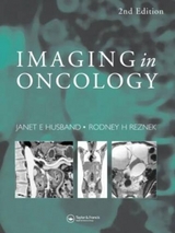
Imaging in Oncology, Second Edition
Taylor & Francis Ltd
978-1-899066-48-3 (ISBN)
- Titel erscheint in neuer Auflage
- Artikel merken
This two-volume set covers image interpretation for tumor staging and follow-up. The editors present imaging findings in different tumor types, their patterns of spread and recurrence, staging considerations, and modern therapeutic approaches. The introductory chapters set the scene with general principles of imaging. The following chapters focus on primary tumor evaluation and metastatic lesions while subspecialty sections discuss pediatric and AIDS-related tumors and detail evaluation of treatment regimes. Although authoritative and comprehensive, Imaging in Oncology distills this exhaustive subject into its essentials.
1. Section I General principles 2. Imaging strategies in oncology: an overview 3. Cancer incidence, trends and survival 4. Staging: purpose and methods 5. Treatment options 6. Section II Primary tumour staging 7. Primary brain tumours 8. Paranasal sinuses 9. Pharynx, tongue, floor of mouth 10. Larynx 11. Thyroid 12. Lung cancer 13. P Armstrong 14. Mediastinal tumours 15. Pleural tumours 16. Oesophagus 17. Stomach and small bowel 18. Colorectal cancer 19. Pancreatic tumours 20. Primary liver tumours 21. Renal tumours 22. Bladder cancer 23. Prostate cancer 24. Testicular tumours 25. Ovarian tumours 26. Uterus and cervical tumours 27. Primary retroperitoneal tumours (including adrenals) 28. Bone tumours 29. Soft tissue tumours 30. Breast cancer 31. Section III Paediatrics 32. Principles of staging paediatric malignancy 33. Nephroblastoma 34. Neuroblastoma 35. Uncommon paediatric tumours 36. Section IV Haematological malignancy 37. Lymphoma 38. Myeloma 39. Leukaemia 40. Section V Metastases 41. Lymph nodes 42. Lung 43. Bone marrow 44. Liver 45. Brain and nervous system 46. Adrenal 47. Peritoneum and omentum 48. Investigation of unknown primary tumours 49. Section VI Treatment evaluation 50. Interventional techniques 51. Imaging for radiotherapy planning 52. Monitoring response and post treatment evaluation 53. Acute complications of treatment 54. Second malignancies 55. Section VII Effects of treatment on normal tissue 56. Lung and chest wall 57. Bone marrow 58. Abdomen and pelvis 59. Section VIII AIDS related tumours 60. Chest 61. Abdomen and pelvis
| Erscheint lt. Verlag | 1.7.1998 |
|---|---|
| Zusatzinfo | 100 Illustrations, black and white |
| Verlagsort | London |
| Sprache | englisch |
| Maße | 219 x 276 mm |
| Gewicht | 4559 g |
| Themenwelt | Medizin / Pharmazie ► Medizinische Fachgebiete ► Onkologie |
| Medizin / Pharmazie ► Medizinische Fachgebiete ► Radiologie / Bildgebende Verfahren | |
| ISBN-10 | 1-899066-48-9 / 1899066489 |
| ISBN-13 | 978-1-899066-48-3 / 9781899066483 |
| Zustand | Neuware |
| Informationen gemäß Produktsicherheitsverordnung (GPSR) | |
| Haben Sie eine Frage zum Produkt? |
aus dem Bereich

