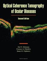
Optical Coherence Tomography of Ocular Diseases
Seiten
2004
|
2nd Revised edition
SLACK Incorporated (Verlag)
978-1-55642-609-4 (ISBN)
SLACK Incorporated (Verlag)
978-1-55642-609-4 (ISBN)
- Titel erscheint in neuer Auflage
- Artikel merken
This work shows the retina in normal and diseased states using the innovative OCT 3000. Topics include macular and other retinal diseases, glaucoma, and a description of OCT technology. An appendix is included for those interested in learning more about the principles of operation.
This work shows the retina in normal and diseased states using the innovative OCT 3000. Topics include macular and other retinal diseases, glaucoma, and a description of OCT technology. An appendix is included for those interested in learning more about the principles of operation of this medical diagnostic imaging technology. The text's primary objective is to illustrate the appearance of the eye in health and disease, comparing conventional clinical technologies with OCT. This method introduces the clinician to the manifestations of disease as elucidated by OCT, while presenting the more familiar funduscopic and fluorescein angiographic appearance side-by-side. The text should provide a clinical reference for the retinal and glaucoma specialist which shows how to utilize and interpret OCT imaging to enhance diagnostic sensitivity and specificity as well as to enhance therapeutic decision-making and monitor the outcome of treatment.
This work shows the retina in normal and diseased states using the innovative OCT 3000. Topics include macular and other retinal diseases, glaucoma, and a description of OCT technology. An appendix is included for those interested in learning more about the principles of operation of this medical diagnostic imaging technology. The text's primary objective is to illustrate the appearance of the eye in health and disease, comparing conventional clinical technologies with OCT. This method introduces the clinician to the manifestations of disease as elucidated by OCT, while presenting the more familiar funduscopic and fluorescein angiographic appearance side-by-side. The text should provide a clinical reference for the retinal and glaucoma specialist which shows how to utilize and interpret OCT imaging to enhance diagnostic sensitivity and specificity as well as to enhance therapeutic decision-making and monitor the outcome of treatment.
| Erscheint lt. Verlag | 31.5.2004 |
|---|---|
| Zusatzinfo | Illustrations (chiefly col.) |
| Sprache | englisch |
| Maße | 111 x 178 mm |
| Themenwelt | Medizin / Pharmazie ► Medizinische Fachgebiete ► Augenheilkunde |
| Medizinische Fachgebiete ► Radiologie / Bildgebende Verfahren ► Computertomographie | |
| ISBN-10 | 1-55642-609-7 / 1556426097 |
| ISBN-13 | 978-1-55642-609-4 / 9781556426094 |
| Zustand | Neuware |
| Haben Sie eine Frage zum Produkt? |
Mehr entdecken
aus dem Bereich
aus dem Bereich


