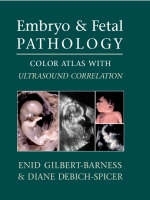
Embryo and Fetal Pathology
Cambridge University Press (Verlag)
978-0-521-82529-0 (ISBN)
- Titel ist leider vergriffen;
keine Neuauflage - Artikel merken
Exhaustively illustrated in color with over 1000 photographs, figures, histopathology slides, and sonographs, this uniquely authoritative atlas provides the clinician with a visual guide to diagnosing congenital anomalies, both common and rare, in every organ system in the human fetus. It covers the full range of embryo and fetal pathology, from point of death, autopsy and ultrasound, through specific syndromes, intrauterine problems, organ and system defects to multiple births and conjoined twins. Gross pathologic findings are correlated with sonographic features in order that the reader may confirm visually the diagnosis of congenital abnormalities for all organ systems. Obstetricians, perinatologists, neonatologists, geneticists, anatomic pathologists, and all practitioners of maternal-fetal medicine will find this atlas an invaluable resource.
Foreword John M. Opitz; Preface; Acknowledgments; 1. The human embryo and embryonic growth disorganization; 2. Late fetal death, stillbirth, and neonatal death; 3. Fetal autopsy; 4. Ultrasound of embryo and fetus: part I. General principles Mark Williams and Kathy B. Porter; Part II Major organ system malformations Mark Williams and Kathy B. Porter; Part III Advances in first trimester ultrasound Susan Guidi; 5. Abnormalities of placenta; 6. Chromosomal abnormalities in the embryo and fetus; 7. Terminology of errors of morphogenesis; 8. Malformation syndromes; 9. Dysplasias; 10. Disruptions and amnion rupture sequence; 11. Intrauterine growth retardation; 12. Fetal hydrops and cystic hygroma; 13. Central nervous system defects; 14. Craniofacial defects; 15. Skeletal abnormalities; 16. Cardiovascular system defects; 17. Respiratory system; 18. Gastrointestinal tract and liver; 19. Genito-urinary system; 20. Congenital tumors; 21. Fetal and neonatal skin disorders; 22. Intrauterine infection; 23. Multiple gestations and conjoined twins; 24. Metabolic diseases; Appendices; Index.
| Erscheint lt. Verlag | 31.5.2004 |
|---|---|
| Co-Autor | Mark Williams, Kathy B. Porter, Susan Guidi |
| Zusatzinfo | 442 Tables, unspecified; 885 Plates, color; 149 Halftones, unspecified; 57 Line drawings, unspecified |
| Verlagsort | Cambridge |
| Sprache | englisch |
| Maße | 290 x 232 mm |
| Gewicht | 2695 g |
| Themenwelt | Medizin / Pharmazie ► Medizinische Fachgebiete ► Gynäkologie / Geburtshilfe |
| Medizin / Pharmazie ► Medizinische Fachgebiete ► Pädiatrie | |
| Studium ► 1. Studienabschnitt (Vorklinik) ► Histologie / Embryologie | |
| Studium ► 2. Studienabschnitt (Klinik) ► Pathologie | |
| ISBN-10 | 0-521-82529-6 / 0521825296 |
| ISBN-13 | 978-0-521-82529-0 / 9780521825290 |
| Zustand | Neuware |
| Haben Sie eine Frage zum Produkt? |
aus dem Bereich


