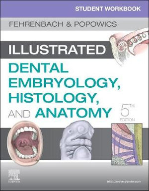
Student Workbook for Illustrated Dental Embryology, Histology and Anatomy
Saunders (Verlag)
978-0-323-63990-3 (ISBN)
- Titel erscheint in neuer Auflage
- Artikel merken
Comprehensive coverage includes all the content needed for an introduction to the developmental, histological, and anatomical foundations of oral health.
Detailed case studies include radiographs, clinical photos, profiles, complaints, health histories, and intraoral examination data, each accompanied by multiple-choice questions, to promote critical thinking skills and prepare students for board examinations.
UNIQUE!
- Guidelines for Tooth Drawing emphasize fundamental principles in tooth design and include detailed instructions, tips, and dimensions for drawing each permanent tooth.
- Glossary exercises include crossword puzzles and word searches for practice and review of key terminology.
- Expert author Margaret Fehrenbach is one of the most trusted names in dental hygiene education.
- Detachable flashcards help students master tooth morphology and tooth numbering, with multiple-angle drawings of a permanent tooth on one side of the flashcard and characteristics of that tooth on the back.
- A logical organization allows students to focus on areas in which they may need more practice, with units on: (1) anatomic and structure identification and labeling, (2) glossary exercises (3) tooth structure, (4) review questions, and (5) case-based application.
- Perforated workbook pages are three-hole-punched so that they easily fit into a binder, and pages can be removed and submitted to instructors for assignments or extra credit.
- NEW! Expanded structure identification exercises include additional developmental, microbiological, and anatomical structures for more practice identifying and labeling various parts or structures.
- NEW! Review questions for each unit provide even more opportunities for content mastery in preparation for classroom or board exams.
- NEW! Additional case studies include questions using the integrated national board format
- Updated! Evidence-based research thoroughly covers infection control of extracted teeth.
- NEW! Occlusal clinical assessment exercises help prepare students for chairside clinical patient care.
STRUCTURE IDENTIFICATION EXERCISES Unit I: Orofacial Structures Unit II: Dental Embryology Unit III: Dental Histology Unit IV: Dental Anatomy
CLINICAL IDENTIFICATION EXERCISES Part 1 - Extraoral Structures Part 2 - Intraoral Structures Part 3 - Tooth Types in Permanent Dentition Part 4 - Permanent Occlusion
GLOSSARY EXERCISES Part 1 - Chapter Word Jumbles Part 2 - Unit Crossword Puzzles Unit I: Orofacial Structures Unit II: Dental Embryology Unit III: Dental Histology Unit IV: Dental Anatomy
TOOTH DRAWING
REVIEW EXERCISES Unit I: Orofacial Structures Unit II: Dental Embryology Unit III: Dental Histology Unit IV: Dental Anatomy
UNIT CASE STUDY EXERCISES Unit I: Orofacial Structures Unit II: Dental Embryology Unit III: Dental Histology Unit IV: Dental Anatomy
| Erscheinungsdatum | 29.11.2019 |
|---|---|
| Zusatzinfo | Approx. 265 illustrations |
| Verlagsort | Philadelphia |
| Sprache | englisch |
| Maße | 216 x 276 mm |
| Gewicht | 610 g |
| Einbandart | kartoniert |
| Themenwelt | Medizin / Pharmazie ► Zahnmedizin ► Studium der Zahnmedizin |
| Schlagworte | Zahnmedizin, Anatomie |
| ISBN-10 | 0-323-63990-9 / 0323639909 |
| ISBN-13 | 978-0-323-63990-3 / 9780323639903 |
| Zustand | Neuware |
| Haben Sie eine Frage zum Produkt? |
aus dem Bereich


