3D Echocardiography of Structural Heart Disease
Springer International Publishing (Verlag)
978-3-319-85303-1 (ISBN)
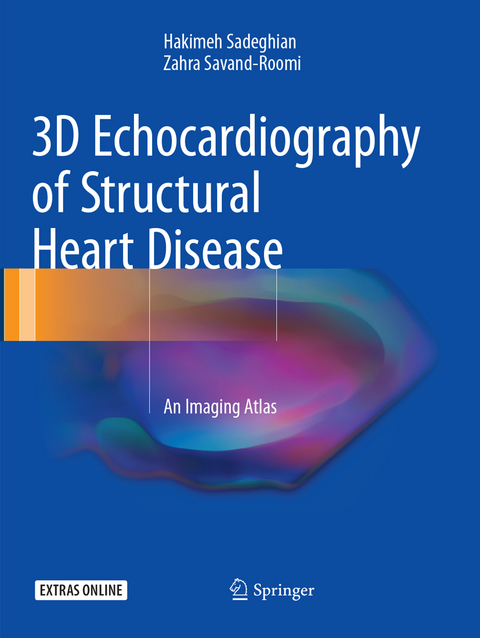
This atlas presents outstanding three-dimensional (3D) echocardiographic images of structural heart diseases, including congenital and valvular diseases and cardiac masses and tumors. The aim is to enable the reader to derive maximum diagnostic and treatment benefit from the modality through optimal image acquisition and interpretation. To this end a wide range of instructive individual cases are depicted, with sequential arrangement of all images and views of diagnostic value, including 3D zoom, full-volume, and live 3D images. For each case, a key lesson is highlighted and attention is drawn to aspects of relevance to diagnosis and treatment. In addition, readers will have online access to echocardiographic video clips for each patient. The closing part of the book examines the role of 3D echocardiography in structural heart disease interventions. The superb quality of the illustrations and the range of cases considered ensure that this atlas will be an excellent visual learning tool and an ideal aid for cardiology residents and fellows in day-to-day clinical practice.
Hakimeh Sadeghian, MD, is Associate Professor of Cardiology and Echocardiography in the Department of Echocardiography, Shariati Hospital, Tehran University of Medical Sciences, Tehran, Iran. Dr. Sadeghian graduated from Tehran University of Medical Sciences in 1992 before specializing in cardiovascular medicine. She later studied in Paris, gaining diplomas in various specialties, including Congenital Heart Disease (Paris University V) and Echocardiography (Paris University XII). Dr. Sadeghian was responsible for founding the Echocardiography Department at Tehran Heart Center, where a range of procedures have been introduced, e.g., transesophageal echocardiography (including during surgery), dobutamine stress echocardiography, tissue Doppler echocardiography, 4D echocardiography, and fetal heart echocardiography. She is co-author (with Zahra Savand-Roomi) of the previous Springer book, Echocardiographic Atlas of Adult Congenital Heart Disease (2015). ; Zahra Savand-Roomi, MD, is a cardiologist in the Department of Echocardiography, Kowsar Hospital, Shiraz, Iran. She graduated from Yazd University of Medical Sciences, Iran, in 2000 before specializing in cardiovascular medicine at Shiraz University of Medical Sciences. Between 2010 and 2012 she studied in Tehran Heart Center, gaining fellowship in Echocardiography (Tehran University of Medical Sciences). Upon returning to Shiraz in 2012 she founded the Echocardiography Department in Kowsar Hospital, where a range of procedures are performed, e.g., transesophageal echocardiography (including during surgery), dobutamine stress echocardiography, tissue Doppler echocardiography and 4D echocardiography. She is co-author (with Hakimeh Sadeghian) of the previous Springer book, Echocardiographic Atlas of Adult Congenital Heart Disease (2015).
Valvular Heart Disease.- Congenital Heart Disease.- Cardiac Masses and Tumors.- Intervention in Structural Heart Disease.
"The intended audience is cardiology residents and fellows in daily clinical practice. ... The book is well organized, easy to read, and uses many high-quality color echocardiographic images to supplement the text. Additionally, readers get online access to echocardiographic video clips for each case. ... This is a valuable tool for those training or practicing in the field of cardiovascular medicine. It is a complete source of information regarding the use of 3D echocardiography in structural heart disease." (Joseph A. Randy Englert, Doody's Book Reviews, March, 2018)
“The intended audience is cardiology residents and fellows in daily clinical practice. … The book is well organized, easy to read, and uses many high‐quality color echocardiographic images to supplement the text. Additionally, readers get online access to echocardiographic video clips for each case. … This is a valuable tool for those training or practicing in the field of cardiovascular medicine. It is a complete source of information regarding the use of 3D echocardiography in structural heart disease.” (Joseph A. Randy Englert, Doody's Book Reviews, March, 2018)
| Erscheint lt. Verlag | 31.8.2018 |
|---|---|
| Zusatzinfo | XIII, 625 p. 24 illus., 12 illus. in color. |
| Verlagsort | Cham |
| Sprache | englisch |
| Maße | 210 x 279 mm |
| Gewicht | 2204 g |
| Themenwelt | Medizinische Fachgebiete ► Innere Medizin ► Kardiologie / Angiologie |
| Medizinische Fachgebiete ► Radiologie / Bildgebende Verfahren ► Radiologie | |
| Schlagworte | 3D echocardiography • Cardiac Masses and Tumors • Cardiology • Congenital Heart Disease • Echocardiographic Video Clips • Structural Heart Disease • Three-dimensional Echocardiographic Images • Valvular Heart Disease |
| ISBN-10 | 3-319-85303-1 / 3319853031 |
| ISBN-13 | 978-3-319-85303-1 / 9783319853031 |
| Zustand | Neuware |
| Haben Sie eine Frage zum Produkt? |
aus dem Bereich


