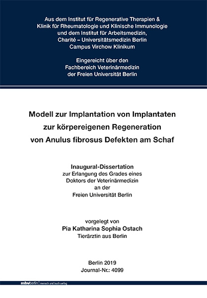
Modell zur Implantation von Implantaten zur körpereigenen Regeneration von Anulus fibrosus Defekten am Schaf
Seiten
2019
|
1. Aufl.
Mensch & Buch (Verlag)
978-3-86387-954-9 (ISBN)
Mensch & Buch (Verlag)
978-3-86387-954-9 (ISBN)
- Keine Verlagsinformationen verfügbar
- Artikel merken
Model for the implantation of implants stimulating the body’s own mechanisms for repair and regeneration of ovine annulus fibrosus defects.
The surgical management of herniated discs often involves removal of the prolapsed tissue and subsequent replacement of the nucleus pulposus. Invasive surgical access via the annulus fibrosus frequently leads to further tissue defects and consequently to additional damage to the existing damage of the annulus fibrosus. Numerous experimental studies on the treatment of degenerative and traumatic changes in intervertebral discs report results of surgical treatment with the aim of repair and regeneration of the nucleus pulposus. The success of these therapeutic methods largely depends on the functional restoration of the annulus fibrosus, since only an intact annulus fibrosus can resist the pressure from inside the intervertebral disc and thus prevent recurrent herniation. Therapeutic methods taking advantage of the regenerative potential of the annulus fibrosus are referred to as Annulus Fibrosus Tissue Engineering. They are based on the implantation of various different scaffolds and cells fostering repair and regeneration.
The aim of this study was to assess the suitability of the model under review to test the suitability of the absorbable implants used for the treatment of annulus fibrosus defects. In the present study, for the first time, an implant in combination with a human recombinant chemokine TECK (CCL25) and in combination with PRP was tested for its bio-compatibility and stability in an in vivo test for annulus fibrosus defect healing in a sheep model. Since the chemokine TECK is a human recombinant chemokine, the effect of this specific chemokine on ovine annulus fibrosus cells was studied in vitro in a chemotaxis assay and also in a 3D cell culture before testing in vivo. In vitro, the human recombinant chemokine TECK was able to dose-dependently induce migration of ovine annulus fibrosus cells, which was significant at concentrations of 750nM and 1000nM. Hence, the receptor ligand of the human recombinant chemokine TECK not only binds to human cells, but also to the chemokine receptor of ovine annulus fibrosus cells. In addition, in 3D cell culture, the ovine annulus fibrosus cells were able to generate proteoglycans in the presence of human recombinant chemokine TECK. In combination with PRP, the ovine annulus fibrosus cells generated almost no proteoglycans. For the first time, the suitability of the human recombinant chemokine TECK for use in animal experiments on sheep could be demonstrated with these in vitro experiments.
In the following animal experiment, 3.5mm x 3.5mm defects were set via a retroperitoneal approach to the annulus fibrosus of the lumbar intervertebral discs of 30 experimental ovine animals. The experimental animals were randomized into 5 groups of 6 (groups A to E). Group A (empty defect), where a defect was set in the annulus fibrosus and subsequently not covered with an implant, was the control group to study the body's own repair and regeneration mechanisms. In groups B to E, the defect was covered with various combinations of implants and therapeutic substances. A macroscopic and histological evaluation was carried out 3 months after surgery.
The assessed model is suitable for testing absorbable implants as this thesis shows, that:
• the sheep was an acceptable model for the study of new surgical therapies with implants for the repair and regeneration of annulus fibrosus defects, since the proportional anatomical conditions are comparable with humans and the same surgical techniques (instruments and implants) were used as would be under clinical conditions for humans. In summary, the model and surgical techniques are transferable to humans. The assessed model is suitable to demonstrate this.
• the retroperitoneal access as well as the defect setting with scalpel and tweezers was reproducible in all animals with increasing efficiency.
• the attachment method with sutures at the four corners of the implant reliably prevented dislocation of the absorbable implants.
• the explantation, anatomical preparation, and preparation of specimens were reproducible.
• histological hematoxylin / eosin staining proved to be a reliable diagnostic tool to study implant bio-degeneration, recurrent herniation rates, and, thus, defect stabilization.
For future use of the assessed model in new studies however, the implant attachment method should be modified to allow for clinically safe, reproducible application and easy integration of the method into existing surgical protocols. A surgical application aid should be used for the challenging application of sutures, since in each instance several attempts to correctly place the implant were required. Also, hematoxylin / eosin staining as used in the assessed model did not allow conclusions as to the quality of the regenerated tissue, and, thus, not as to the effectiveness of the applied therapeutic substances on tissue repair and regeneration. Only quantitative effects could be assessed. The hypothesis, whether human recombinant chemokine TECK (CCL25), in addition to the demonstrated in vitro effect, also has an in vivo effect on defect healing of ovine annulus fibrosus cells, could not be assessed with the methods under review. Hence, for future use of the model, defects of equal size should be set in all animals, and the study of the defect content should also be in the focus of further research. Since the model is suitable in principle, with defects set of equal size, a classification system for quantitative analysis of tissue repair and regeneration between groups of animals as well as relative to the chemokine TECK used can be developed. Die operative Versorgung von Bandscheibenvorfällen umfasst häufig die Entfernung des prolabierten Gewebes und den sich anschließenden Ersatz des Nucleus pulposus. Durch den hierzu gesetzten Zugang über den Anulus fibrosus kommt es, neben dem schon bestehenden Schaden im Anulus fibrosus, häufig zu einer weiteren Defektsetzung und damit zur weiteren Schädigung des Anulus fibrosus. Zahlreiche experimentelle Studien über die Therapie von Bandscheibenveränderungen berichten von Ergebnissen zur regenerativen und restaurativen Versorgung des Nucleus pulposus. Der Erfolg dieser Therapiemethoden hängt maßgeblich von der funktionellen Wiederherstellung des Anulus fibrosus ab, da nur ein intakter Anulus fibrosus den Druckverhältnissen innerhalb der Bandscheibe standhalten und damit eine erneute Herniation verhindern kann. Therapieansätze, die sich mit dem Regenerationspotential des Anulus fibrosus beschäftigen, werden unter dem Begriff Anulus fibrosus Tissue Engineering zusammengefasst. Sie basieren auf der Nutzung von verschiedenen Gerüsten (Scaffolds) und von Zellen zur Regeneration. Das Ziel dieser Arbeit war zu bewerten, ob das genutzte Modell geeignet ist, diese Implantate zur Therapie von Anulus fibrosus Defekten auf ihre Eignung zu prüfen.
Als Prüfpräparat wurde in der vorliegenden Arbeit erstmals ein Implantat in Kombination mit einem human rekombinanten Chemokin TECK (CCL25) und in Kombination mit PRP in einem in vivo Versuch zur Anulus fibrosus Defektheilung im Schafmodell auf seine Bioverträglichkeit und seine Stabilität getestet. Da es sich bei dem Chemokin TECK um ein human rekombinantes Chemokin handelt, wurde vorab die Wirkung des Chemokins auf ovine Anulus fibrosus Zellen in vitro in einem Chemotaxisassay und in einer 3D-Zellkultur untersucht. In vitro konnte das human rekombinante Chemokin TECK die ovinen Anulus fibrosus Zellen dosisabhängig zur Migration anregen und dies in einer Konzentration von 750nM und 1000nM signifikant. Der Rezeptorligand des human rekombinanten Chemokins TECK geht demnach nicht nur mit humanen Zellen eine Bindung ein, sondern auch mit dem Chemokinrezeptor oviner Anulus fibrosus Zellen. Zusätzlich konnten die ovinen Anulus fibrosus Zellen in der 3DZellkultur in Anwesenheit des human rekombinanten Chemokins TECK Proteoglykane bilden. In Kombination mit dem verwendeten PRP bildeten die ovinen Anulus fibrosus Zellen nahezu keine Proteoglykane. Die Grundlage für die sich anschließende Verwendung des human rekombinaten Chemokins TECK, im tierexperimentellen Versuch am Schaf, konnte mit diesen in vitro Versuchen erstmals nachgewiesen werden.
Im folgenden Tierversuch wurden über einen retroperitonealen Zugang 3,5mm x 3,5mm große Defekte in den Anulus fibrosus der Lendenwirbelsäulenbandscheibe von 30 ovinen Versuchstieren appliziert. Hierbei wurden die Versuchstiere randomisiert in 5 Gruppen à 6 Tiere (Gruppen A bis E) eingeteilt. Gruppe A (Leerdefekt) stellte die Kontrollgruppe zur Überprüfung der körpereigenen Reaktions- und Regenerationsmechanismen dar, bei der ein Defekt in den Anulus fibrosus gesetzt und anschließend nicht mit einem Implantat verschlossen wurde. Bei den Gruppen B bis E wurde der Defekt mit verschiedenen Prüfpräparaten und Wirkstoffkombinationen gedeckt. Eine makroskopische und histologische Auswertung wurde nach 3 Monaten Standzeit der Tiere durchgeführt.
Das beschriebene Modell ist geeignet, Implantate auf ihre Eignung zu prüfen, da:
• das ovine Versuchstier ein akzeptables Modell für die Untersuchung neuer Therapien in Form von Implantaten zur Anulus fibrosus Defektheilung darstellte, weil vergleichbare anatomische Größenverhältnisse zum Menschen vorliegen und bei der verwendeten Operationstechnik (Instrumente und Implantate) dieselben Techniken zum Einsatz kamen, wie sie auch unter klinischen Gegebenheiten am Mensch genutzt werden sollen. In Summe sind das Modell und die Operationstechnik damit auf den Menschen übertragbar und das Modell geeignet, dies zu zeigen.
• der genutzte retroperitoneale Zugang, sowie die Defektsetzung mit Skalpell und Pinzette sich bei allen Tieren mit steigender Effizienz reproduzierbar durchführen ließen.
• das genutzte Befestigungsverfahren in Form von Nahtmaterial an den vier Ecken des Implantats, eine Dislokation der Implantate sicher verhindern konnte.
• die Explantation, die Präparation und die Herstellung der Präparate reproduzierbar durchführbar waren.
• die genutzte histologische Hämatoxylin/Eosin-Färbung sich als zuverlässiges Diagnostikum erwies, um die Biodegeneration des Implantates, die Herniationsraten und damit die Defektstabilität zu evaluieren.
Bei erneuter Anwendung des beschriebenen Modells, sollte jedoch das Befestigungsverfahren modifiziert werden, um eine klinische, sichere, reproduzierbare Anwendung und die einfache Integration des Verfahrens in die bestehenden chirurgischen Arbeitsabläufe zukünftiger Studien zu ermöglichen. Für das genutzte, anspruchsvolle Befestigungsverfahren sollte ein Applikationsinstrument genutzt werden, da jeweils mehrere Versuche zur korrekten Platzierung des Implantats notwendig waren. Die im beschriebenen Modell genutzte Hämatoxylin/Eosin-Färbung ließ außerdem keine Rückschlüsse auf die Qualität des entstandenen Regeneratgewebes zu und somit auch nicht auf die Wirkung der eingesetzten Stoffe auf die Geweberegeneration, sondern lediglich die Quantität. Die Hypothese, ob das eingesetzte Chemokin TECK neben der in vitro nachgewiesenen Wirkung auf die ovinen Anulus fibrosus Zellen, auch in vivo einen Einfluss auf die Defektheilung hatte, konnte mit den verwendeten Methoden nicht beurteilt werden. Somit sollte bei einer erneuten Nutzung des Modells der Defekt in bei allen Tieren in gleicher Größe gesetzt werden und anschließend auch die Untersuchung des Defektinhaltes im Fokus weiterer Forschungsarbeiten stehen. Da das Modell grundsätzlich geeignet ist, kann bei gleicher Defektgröße zukünftig ein Klassifikationssystem einen qualitativen Vergleich der Geweberegeneration der Versuchstiergruppen untereinander und damit auch eine Auswertung in Bezug auf das verwendete Chemokin TECK zulassen.
| Erscheinungsdatum | 06.03.2019 |
|---|---|
| Verlagsort | Berlin |
| Sprache | deutsch |
| Themenwelt | Medizinische Fachgebiete ► Innere Medizin ► Rheumatologie |
| Veterinärmedizin ► Vorklinik | |
| Veterinärmedizin ► Großtier ► Schaf / Ziege | |
| Schlagworte | Animal Models • anulus fibrosus (MeSH) • Cell Cultures • Healing • Implantation • intervertebral discs • Regeneration • Schafe • sheep, • Tiermodelle |
| ISBN-10 | 3-86387-954-6 / 3863879546 |
| ISBN-13 | 978-3-86387-954-9 / 9783863879549 |
| Zustand | Neuware |
| Informationen gemäß Produktsicherheitsverordnung (GPSR) | |
| Haben Sie eine Frage zum Produkt? |
Mehr entdecken
aus dem Bereich
aus dem Bereich
Das Wichtigste für Klinik und Praxis
Buch | Softcover (2022)
Urban & Fischer in Elsevier (Verlag)
39,00 €
Information für Erkrankte, Angehörige und Betreuende
Buch | Softcover (2023)
Springer (Verlag)
22,99 €


