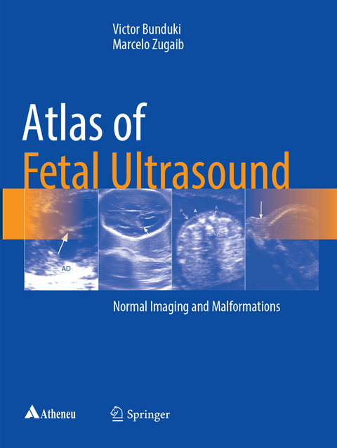
Atlas of Fetal Ultrasound
Springer International Publishing (Verlag)
978-3-319-85485-4 (ISBN)
The present book is an illustrated guide on fetal medicine, including a wealth of normal and pathological/malformations ultrasound images, throughout the whole pregnancy. Thus, the book intends to fill two gaps in once:
- The lack of a material discussing the basic principles of fetal ultrasound, which are the basement for a more efficient learning in fetal medicine.
- . The need for a thorough approach in fetal medicine, presenting both normal and pathological imaging, allowing a detailed evaluation of clinical conditions of importance in prenatal care and follow up.
The Atlas of Ultrasound Imaging is an up to date guide to all obstetricians, gynecologists and pediatricians who intend to upgrade their knowledge in fetal medicine, as well as to any other professional, professor, student or research interested in fetal ultrasound.
Victor Bunduki University of Sao Paulo Medical School Sao Paulo, Brazil MD by the University of Sao Paulo, Brazil (USP - 1988), Master and PhD in Obstetrics and Gynecology by USP (1995 and 1998). Medical residency by USP (1991) and by Universite Paris Descartes, France (1992). Associate professor at USP Medical School (FMUSP). Effective member of the International Fetal Medicine and Surgery Society since 1992. Marcelo Zugaib Universityof Sao Paulo Medical School Sao Paulo, Brazil Medical residency in obstetrics and gynecology by University of Sao Paulo (USP) Medical School and fellowship by the University of California-Los Angeles, USA (UCLA). Master and PhD by USP. Full professor of Obstetrics at USP Medical School.
Chapter 1 Normal Fetal Morphological Ultrasound.- Chapter 2 The Fetus at the First Trimester.- Chapter 3 Central Nervous System Abnormalities.- Chapter 4 Neural Tube Defects.- Chapter 5 Ultrasound Evaluation of Fetal Face.- Chapter 6 The Fetal Heart Standpoint of General Sonographer.- Chapter 7 Fetal Echocardiography.- Chapter 8 Noncardiac Thoracic Malformations.- Chapter 9 The Fetal Abdomen.- Chapter 10 Urinary Tract.- Chapter 11 Genitals.- Chapter 12 Skeletal and Limbs.- Chapter 13 Soft Tissues.- Chapter 14 Umbilical Cord and Placenta.- Chapter 15 Ultrasound in Fetal Infections.- Chapter 16 Multiple Gestation.- Chapter17 Fetal Aneuploidies.- Chapter 18 Invasive Procedures in Fetal Medicine.- Chapter 19 Three-Dimensional Ultrasound.- Chapter 20 Doppler Ultrasound in Obstetrics.
| Erscheint lt. Verlag | 30.8.2018 |
|---|---|
| Zusatzinfo | IX, 261 p. 966 illus., 122 illus. in color. |
| Verlagsort | Cham |
| Sprache | englisch |
| Maße | 210 x 279 mm |
| Gewicht | 963 g |
| Themenwelt | Medizin / Pharmazie ► Gesundheitsfachberufe ► Hebamme / Entbindungspfleger |
| Medizin / Pharmazie ► Medizinische Fachgebiete ► Gynäkologie / Geburtshilfe | |
| Medizinische Fachgebiete ► Radiologie / Bildgebende Verfahren ► Radiologie | |
| Medizinische Fachgebiete ► Radiologie / Bildgebende Verfahren ► Sonographie / Echokardiographie | |
| Schlagworte | Development • diagnostic radiology • Fetal imaging • Fetal Medicine • Imaging • Pregnancy • ultrasonography |
| ISBN-10 | 3-319-85485-2 / 3319854852 |
| ISBN-13 | 978-3-319-85485-4 / 9783319854854 |
| Zustand | Neuware |
| Informationen gemäß Produktsicherheitsverordnung (GPSR) | |
| Haben Sie eine Frage zum Produkt? |
aus dem Bereich


