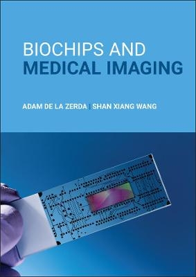
Biochips and Medical Imaging
John Wiley & Sons Inc (Verlag)
978-1-118-91050-4 (ISBN)
Biochips and Medical Imaging is designed as a professional resource, covering recent biochip and medical imaging developments. Within the text, the authors encourage uniting aspects of engineering, biology, and medicine to facilitate advancements in the field of molecular diagnostics and imaging.
Biochips are microchips for efficiently screening biological analytes. This book aims at presenting information on the state-of-the-art and emerging biosensors, biochips, and imaging devices of the body's systems, including the endocrine, circulatory, and immune systems.
Medical diagnostics includes biochips (in-vitro diagnostics) and medical and molecular imaging (in-vivo imaging). Biochips and Medical Imaging explores the role of in-vitro and in-vivo diagnostics. It enables an instructor to share in-depth examples of the use of biochips in diagnosing cancer and cardiovascular diseases.
Provides real-life knowledge on biochips and medical imaging, written by leading researchers
Serves as a resource for professionals working in the biochip or imaging fields
Features an accessible approach for anyone interested in biochips and their applications
Readers of Biochips and Medical Imaging can expand their knowledge of medical technology, even if they have no biological knowledge and a limited math background. With its focus on important developments, this book is sure to also capture the interest of bioengineering and biomaterials scientists, structural biologists, electrical engineers, and nanotechnologists.
ADAM DE LA ZERDA is an Associate Professor in the departments of Structural Biology and Electrical Engineering at Stanford University and a Chan Zuckerberg Investigator. Dr. de la Zerda has received numerous awards for his research including the Forbes Magazine 30-under-30 in Science & Healthcare and published over 30 papers in leading journals. He holds a number of patents and is the founder of the medical diagnostics company Visby Medical. SHAN XIANG WANG is the Leland T. Edwards Professor in the School of Engineering, Stanford University. He currently serves as the Director of the Stanford Center for Magnetic Nanotechnology and a Professor of Materials Science & Engineering, jointly of Electrical Engineering, and by courtesy of Radiology . He has published over 300 papers, and holds 66 patents (issued and pending). Dr. Wang was an inaugural Frederick Terman Faculty Fellow at Stanford University (1994–1997), an IEEE Magnetics Society Distinguished Lecturer (2001–2002), and elected an IEEE Fellow (2009) and American Physical Society (APS) Fellow (2012).
Foreword xvii
Preface xix
Acknowledgments xxi
1 Cell Biology 1
1.1 Cell Biology Introduction 1
1.2 Cell Structure 1
1.3 Cell Membrane 2
1.4 Proteins 2
1.5 Cytoplasm and Organelles 3
1.6 Nucleus 6
1.7 Nucleic Acids (DNA and RNA) 8
1.8 Central Dogma and Recent Revisions 10
1.9 Mutations 14
1.10 Cell Cycle 14
1.11 Additional Information 17
2 Biological Lab Techniques 27
2.1 Overview 27
2.2 Beer Lambert's Law 27
2.3 DNA Lab Techniques 28
2.4 Additional Information 38
3 Human Physiology 47
3.1 Overview 47
3.2 Nervous System 47
3.3 Circulatory System 68
3.4 Endocrine System 74
3.5 Lymphatic System 83
3.6 Immune System 85
4 Cancer 103
4.1 Epidemiology (Statistics) 103
4.2 What Causes Cancer 104
4.3 Oncogenesis (Cancer Development) 106
4.4 The Six Hallmarks of Cancer 109
4.5 Conclusion 118
5 Cardiovascular Diseases (CVDs) 123
5.1 Epidemiology and Introduction 123
5.2 Types of CVD 125
5.3 Diagnosis of CVDs 130
5.4 Treatment of CVDs 135
5.5 Conclusion 138
6 DNA Chips and Sequencing 143
6.1 Introduction to DNA Chips and PCR 143
6.2 Polymerase Chain Reaction (PCR) 143
6.3 DNA and RNA Chip Technology 147
6.4 DNA Sequencing 155
6.5 Conclusion 156
6.6 Additional Information 156
7 Next-Generation Sequencing and FET-Based Biochips 161
7.1 Introduction to Next-Generation Sequencing 161
7.2 Optical-Based Methods 162
7.3 Electronic-Based Methods 165
7.4 Conclusion 172
8 Protein Assays and Chips 179
8.1 Introduction 179
8.2 ELISA 179
8.3 Protein Arrays 183
8.4 Conclusion 190
8.5 Additional Information 190
9 Label-Free Affinity-Based Biosensors 197
9.1 Introduction 197
9.2 Surface Plasmon Resonance (SPR) Sensor 197
9.3 Nanowire Field-Effect (FET) Sensors 203
9.4 Cantilever Sensors 204
9.5 Electrochemical Sensors 205
9.6 Multiplex Detection of Polymicrobial UTI (Urinary Tract Infection) 207
9.7 Conclusion 211
10 Magneto-Nanosensor Biochips 215
10.1 Magnetism Overview 215
10.2 GMR Magneto-Nanosensor Biochips 216
10.3 Point-of-Care Testing 223
10.4 Non-GMR Magnetic Nanobiosensors 228
10.5 Conclusion 231
11 Microfluidic Chips for Capturing Circulating Tumor Cells 235
11.1 Circulating Tumor Cells 235
11.2 Identifying CTC and WBC by 3-Color Staining 235
11.3 Fluorescence-Activated Cell Sorting (FACS) 236
11.4 Magnetically Activated Cell Sorting (MACS) 237
11.5 Magnetic Separation Devices 238
11.6 CTC Enrichment By Size Filtering 243
11.7 CTC-CHIP (HARVARD UNIVERSITY) 243
11.8 Clinical Utility From CTCs 245
11.9 Conclusion 247
12 Molecular Diagnostics 251
12.1 Molecular Diagnostics (Dx) 251
12.2 Molecular Diagnostics for Cancer 251
12.3 Important Concepts in Diagnostics 254
12.4 Conclusion 261
12.5 Additional Information 261
13 Magnetic Resonance Imaging 271
13.1 Medical Imaging -- Categorization 271
13.2 Overview For Imaging Section 271
13.3 MRI: Past, Present, and Future 273
13.4 Physics of MRI Overview 274
13.5 Physics of MRI 274
13.6 Image Acquisition in MRI 279
13.7 MRI Contrast Agents 282
13.8 Conclusion 287
14 Radionuclide Imaging 295
14.1 Radioactivity 295
14.2 Basics of Positron Emission Tomography (PET) 299
14.3 Single-Photon Emission Computer Tomography (SPECT) 303
14.4 Contrast and Imaging Agents 306
14.5 Conclusion 312
15 Fluorescence and Raman Imaging 317
15.1 Introduction to Optical Imaging 317
15.2 Photon/Tissue Interaction 317
15.3 Fluorescence Imaging 320
15.4 Raman Imaging 328
15.5 Fluorescence Imaging vs. Raman Imaging 331
15.6 Conclusion 332
16 Optical Coherence Tomography 337
16.1 Introduction 337
16.2 Applications of OCT 346
16.3 Contrast Enhancement 351
16.4 Conclusion 359
17 Photoacoustic Imaging 363
17.1 Photoacoustic Effect 363
17.2 The Thermal and Stress Confinements 364
17.3 Photoacoustic Imaging 365
17.4 Contrast Agents 367
17.5 Conclusion 373
18 Imaging Controls and Concepts 377
18.1 Controls 377
18.2 Imaging Concepts 382
18.3 Clinical Translation 386
18.4 Conclusion 390
Problems 390
References 394
Further
Reading 394
Index 395
| Erscheinungsdatum | 17.08.2022 |
|---|---|
| Verlagsort | New York |
| Sprache | englisch |
| Maße | 10 x 10 mm |
| Gewicht | 454 g |
| Themenwelt | Medizin / Pharmazie ► Medizinische Fachgebiete |
| Medizin / Pharmazie ► Physiotherapie / Ergotherapie ► Orthopädie | |
| Naturwissenschaften ► Biologie | |
| Technik ► Maschinenbau | |
| Technik ► Medizintechnik | |
| Technik ► Umwelttechnik / Biotechnologie | |
| ISBN-10 | 1-118-91050-8 / 1118910508 |
| ISBN-13 | 978-1-118-91050-4 / 9781118910504 |
| Zustand | Neuware |
| Informationen gemäß Produktsicherheitsverordnung (GPSR) | |
| Haben Sie eine Frage zum Produkt? |
aus dem Bereich


