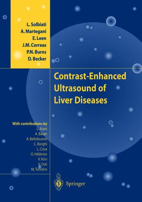
Contrast-Enhanced Ultrasound of Liver Diseases
Springer Verlag
978-88-470-2168-6 (ISBN)
- Titel wird leider nicht erscheinen
- Artikel merken
1 Contrast Ultrasound Technology.- 1.1 Introduction: The Need for Bubble-Specific Imaging.- 1.1.1 Bubble Behaviour and Incident Pressure.- 1.1.2 The Mechanical Index (MI).- 1.2 Nonlinear Backscatter: Harmonic Imaging.- 1.2.1 Harmonic B-Mode Imaging.- 1.2.2 Harmonc Doppler.- 1.2.3 Harmonic Power Doppler Imaging.- 1.2.4 Tissue Harmonic Imaging.- 1.2.5 Pulse Inversion Imaging.- 1.2.6 Power Pulse Inversion Imaging.- 1.3 Transient Disruption: Intermittent Imaging.- 1.3.1 Triggered Imaging.- 1.3.2 Intermittent Harmonic Power Doppler for Perfusion Imaging.- 1.4 Summary.- 1.5 Safety Considerations.- 1.6 Conclusion.- References.- 2 Ultrasound Contrast Agents.- 2.1 Introduction.- 2.2 Basic Physics and Pharmacology.- 2.3 Commercial Ultrasound Contrast Media.- 2.3.1 Albunex (TM).- 2.3.20ptison (TM).- 2.3.3 Levovist (R).- 2.3.4 Sono Vue (R).- 2.3.5 Definity (TM).- 2.3.6 Imagent (TM).- 2.4 Future Developments in Ultrasound Contrast Media.- References.- 3 Liver Physiology.- 3.1 Introduction.- 3.2 Anatomy.- 3.3 Liver Lobule Structure.- 3.3.1 Liver Cell Plate.- 3.3.2 Nonparenchymal Liver Cells.- 3.3.3 Sinusoidal Endothelial Cells.- 3.3.4 Kupffer Cells.- 3.3.5 Hepatic Stellate Cells.- 3.3.6 Fibrosis and Hepatic Stellate Cells.- 3.3.7 Pit Cells.- 3.4 Hepatocyte Transport.- 3.4.1 Electrolyte and Solute Transport.- 3.4.2 Bile Formation.- 3.5 Functional Organization of the Liver Cell Plate.- 3.5.1 Limiting Plate.- 3.5.2 Hepatocyte Heterogeneity Along the Liver Cell Plate.- 3.5.3 Carbohydrate Metabolism.- 3.5.4 Fatty Acid Metabolism.- 3.5.5 Amino Acid and Ammonia Metabolism.- 3.5.6 Regulation of Hepatocyte Heterogenicity.- 3.5.7 Physiologic Significance of Hepatocyte Heterogenicity.- References.- 4 Macro- and Microcirculation of Focal Liver Lesions.- 4.1 Traumatic Abdominal Lesions.- 4.1.1 Introduction.- 4.1.2 Ultrasound and Abdominal Trauma.- 4.1.3 Contrast-Enhanced Ultrasound Image Gallery.- 4.1.4 Conclusions.- References.- 4.2 Hepatic Hemangiomas.- 4.2.1 Anatomical and Structural Features.- 4.2.2 Diagnostic Imaging.- 4.2.3 Integrated Imaging.- 4.2.4 Contrast-Enhanced Ultrasound Imaging.- 4.2.5 Differential Diagnostic Problems1.- References.- 4.3 Hepatic Adenoma.- References.- 4.4 Macro-and Microcirculation of Focal Nodular Hyperplasia.- References.- 4.5 Other Benign Lesions and Pseudo lesions.- 4.5.1 Inflammatory Pseudotumor of the Liver.- 4.5.2 Hepatic Tuberculoma.- 4.5.3 Focal Fatty Changes.- 4.5.4 Skip Areas in Diffuse Steatosis.- References.- 4.6 Hepatocellular Carcinoma and Dysplastic Lesions.- 4.6.1 Introduction and Epidemiology.- 4.6.2 Pathology and Hepatocarcinogenesis.- 4.6.3 Unenhanced Sonography.- 4.6.4 Contrast-Enhanced Ultrasound.- References.- 4.7 Liver Metastases.- 4.7.1 Anatomical and Structural Features.- 4.7.2 Diagnostic Imaging.- 4.7.3 Integrated Imaging.- 4.7.4 Contrast-Enhanced Ultrasound Imaging.- 4.7.5 Differential Diagnostic Problems.- References.- 4.8 Other Malignancies.- 4.8.1 Peripheral Cholangiocarcinoma.- 4.8.2 Hepatic Sarcoma.- 4.8.3 Hepatic Lymphoma.- References.- 5 Medical Needs.- 5.1 Introduction.- 5.2 Incidental Lesions in Normal Livers.- 5.3 Screening and Surveillance for Hepatocellular Carcinoma.- 5.4 Staging and Follow-up of Cancer Patients1.- References.- 6 Guidance and Control of Percutaneous Treatments.- 6.1 Introduction.- 6.2 Fundamentals and Pathology of Ablative Treatments.- 6.3 Role of Diagnostic Imaging.- 6.4 Detection of Lesions and Selection of Patients.- 6.5 RF Procedures and Targeting of Lesions.- 6.6 Assessment of Therapeutic Response.- References.- Hands-on Contrast Ultrasound.- 7.1 Tips and Tricks.- 7.1.1 Organization of Contrast-Enhanced US.- 7.1.2 Intravenous Access.- 7.1.3 Preparation of the USCA.- 7.1.4 The USCAs.- 7.1.5 Scanning Techniques and Settings.- 7.1.6 Liver CEUS using Levovist (R).- 7.1.7 Liver CEUS using SonoVue (R).- 7.2 Limitations.- 7.3 Artifacts and Pitfalls.- 7.3.1 Heterogeneous Enhancement.
| Erscheinungsdatum | 20.12.2018 |
|---|---|
| Co-Autor | M. Tonolini, L. Aiani |
| Zusatzinfo | XII, 123 p. |
| Verlagsort | Milan |
| Sprache | englisch |
| Maße | 216 x 280 mm |
| Themenwelt | Medizinische Fachgebiete ► Innere Medizin ► Gastroenterologie |
| Medizinische Fachgebiete ► Radiologie / Bildgebende Verfahren ► Radiologie | |
| Medizinische Fachgebiete ► Radiologie / Bildgebende Verfahren ► Sonographie / Echokardiographie | |
| Schlagworte | carcinoma • Cell • Contrast-enhanced sonography • focal liver lesions • hepatocellular carcinoma • Imaging • Liver • liver disease • Metastasis • pharmacology • Radiofrequency ablation • sonography • Ultrasound |
| ISBN-10 | 88-470-2168-5 / 8847021685 |
| ISBN-13 | 978-88-470-2168-6 / 9788847021686 |
| Zustand | Neuware |
| Informationen gemäß Produktsicherheitsverordnung (GPSR) | |
| Haben Sie eine Frage zum Produkt? |
aus dem Bereich


