Ultrastructure of the Digestive Tract -

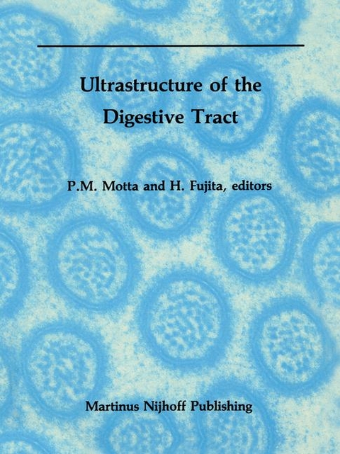
Ultrastructure of the Digestive Tract

P. Motta, H. Fujita (Herausgeber)

Buch | Softcover
262 Seiten
1989 | Softcover reprint of the original 1st ed. 1988
Springer-Verlag New York Inc.
978-1-4612-9229-6 (ISBN)
192,59 inkl. MwSt
  • Titel wird leider nicht erscheinen
  • Artikel merken
When established four years ago, the scope of this international series in electron microscopy essentially was to provide an opportunity for the pUblication of selected review contributions by specialists in ultrastructural research. Previous volumes presented over the last three years have focused on special topics of present interest in ~'ontemporary biomedicine such as endocrine cells, reproduction, and connective tissues. In these fielCls, in fact, integrated methods of electron microscopy have contributed much to generate new ideas and concepts of general value in both basic and clinical applications. The Ultrastructure of the Digestive Tract basically follows the same guidelines and style of the other books in the series and is an invited collection of selected contributions of authors from various laboratories active in the field of electron microscopy. Therefore, although the various chapters consist of individual topics, they nevertheless should be considered as interrelated contributions of specific subjects in the field. The idea was to have critical reviews of aspects previously published elsewhere by experts in the field who, as a rule, include other relevant information in their articles in order to update and enrich the subject. This book contains fifteen chapters by renowned electron microscopists. Each chapter, according to the policy of the editors, reviews a particular topic in great detail, providing updated information, study methods and results, authors' ideas on future investigative approaches, and possible guidelines for forthcoming work. We hope that this book will be useful to cell biologists, morphologists, physiologists, and pathologists.

1. Morphological changes in the developing alimentary canal: A review by scanning electron microscopy.- 2. The esophagus: Normal ultrastructure and pathological patterns.- 3. Fine structure of gastric glands.- 4. Ultrastructural aspects of the turnover of stomach mucosal epithelium.- 5. Biology of the duodenal (Brunner's) glands.- 6. Jejunum and villi: Structural basis of intestinal absorption.- 7. Ultrastructure of the normal and diseased appendix and cecum.- 8. The colon: Normal ultrastructure and pathological patterns.- 9. Ultrastructure of the rectum with special emphasis on tumors.- 10. The musculature and innervation of the alimentary canal.- 11. The lymphoid system and immunological defense of the digestive tract.- 12. Microvascularization of the alimentary canal as studied by scanning electron microscopy of corrosion casts.- 13. The endocrine cell system of the digestive tract.- 14. Morphology and porosity of the alimentary epithelial basal lamina.- 15. The peritoneum.

Erscheinungsdatum
Reihe/Serie Electron Microscopy in Biology and Medicine ; 4
Zusatzinfo XII, 262 p.
Verlagsort New York, NY
Sprache englisch
Maße 216 x 280 mm
Themenwelt Studium 1. Studienabschnitt (Vorklinik) Anatomie / Neuroanatomie
Schlagworte Absorption • Biology • biomedicine • Cell • Cells • clinical application • Esophagus • Medicine • Microscopy • Morphology • Production • Reproduction • Research • Stomach • tissue
ISBN-10 1-4612-9229-8 / 1461292298
ISBN-13 978-1-4612-9229-6 / 9781461292296
Zustand Neuware
Haben Sie eine Frage zum Produkt?
Mehr entdecken
aus dem Bereich
Struktur und Funktion

von Martin Trepel

Buch | Softcover (2021)
Urban & Fischer in Elsevier (Verlag)
44,00