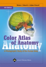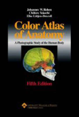
Color Atlas of Anatomy
Lippincott Williams and Wilkins (Verlag)
978-0-7817-3194-2 (ISBN)
- Titel erscheint in neuer Auflage
- Artikel merken
The on-going core of this atlas is its standard of realistic illustrations that portray anatomical relationships. Photographs of actual cadaver dissections along with numerous schematic drawings aid the student in anatomic orientation. Chapters are organized by region, in order of a typical dissection. Each chapter contains two sections: a description and illustration of organs, and a depiction of those organs within the regional anatomy. New to this edition is an increase of MRI pictures, approximately 30 schematic drawings made even more precise, and an updated text where appropriate. This is a Brandon-Hill recommended title.
1. General Anatomy Organization of the Human Body Skeleton of the Human Body Ossification of the Bones Bone Structure Arthrology Principal Joints (Immovable) Synovial Joints (Movable) Shapes of Muscles Muscles, General Myology Organization of the Nervous System Organization of the Circulatory System Organization of the Lymphatic System 2. Head and Neck Bones of the Skull Disarticulated Skull I Disarticulated Skull II Disarticulated Skull III (Cranial Bones) Calvaria Base of the Skull from above Median Section through the Skull Disarticulated Skull IV (Facial Skeleton) Base of the Skull from below Mandible and Dental Arch Temporomandibular Joint Muscles of the Temporomandibular Joint Faci Muscles Supra- and Infrahyoid Muscles Coronal Section Cranial Nerves -Nerves of the Orbit -Trigerninal Nerve -Facial Nerve Glossopharyngeal, Vagus and Hypoglossal Nerve Superficial Region of the Face Retromandibular Region Para- and Retropharyngeal Region Skull of the Newborn Scalp Meninges -Dura Mater and Dural Venous Sinuses -Pia Mater and Arachnoid Brain -Sagittal Sections -ArteriesandVeins -Lobes of the Cerebrum -Lobes of the Cerebeilnin -Dissection -Limbic System -Hypothalamus Subeortical Nuclei VentricularSystem BrainStem Cross Sections Horizontal Sections Auditory and Vestibular Apparatus -Middle Ear -Auditory Ossicles Internal Ear Labyrinth Acoustic Pathway Visual Apparatus and Orbit Eyeball Vessels of the Eye Extraocular Muscles Visual Pathway Layers of the Orbit Lacritnal Apparatus and Lids Nasal Septum Nasal Cavity and Paranasal Sinuses -Nerves and Arteries -Sections Oral Cavity -Subrnandibular Triangle -Salivary Glands General Organization of the Neck Muscles of the Neck Larynx -Muscles -Vocal Ligament -Innervation Larynx and Oral Cavity Pharynx -Muscles Arteries Veins Lymph Vessels Anterior Aspect of the Neck Posterior Triangle Lateral Aspect of the Neck Cervical and Brachial Plexus Sections through the Neck 3. Trunk Skeleton Vertebrae Vertebral Column and Thorax Ribs and Vertebra Joints Costovertebral Joints Ligaments Muscles of the Thorax Thoracic Wall Thoracic and Abdominal Wall 4. Thoracic Organs Position of the Thoracic Organs Respiratory System Projections of the Lungs and Pleura Lungs Bronchopulmonary Segments Heart Myocardium Valves Function Conducting System Vessels Regional Anatomy of the Thoracic Organs Thymus Heart Pericardiwn Posterior Mediastinum Mediastinal Organs Diaphragm Coronal Sections through the Thorax Horizontal Sections through the Thorax Fetal Circulatory System Mammary Gland 5. Abdominal Organs Digestive System Anterior Abdominal Wall Stomach Pancreas and Bile Ducts Liver Spleen and Portal Circulation Vessels of the Abdominal Organs Dissection of Abdominal Organs Mesenteric Arteries Abdominal Cavity Upper Abdominal Organs Lesser Sac Celiac Trunk Posterior Abdominal Wall Cavity Pancreas Pancreas and Spleen Root of the Mesentery Diaphragms and Peritoneal Recesses Sections through the Abdominal Cavity 6. Retroperitoneal Orga
| Erscheint lt. Verlag | 15.3.2002 |
|---|---|
| Zusatzinfo | 1158 |
| Verlagsort | Philadelphia |
| Sprache | englisch |
| Maße | 216 x 299 mm |
| Gewicht | 2397 g |
| Themenwelt | Studium ► 1. Studienabschnitt (Vorklinik) ► Anatomie / Neuroanatomie |
| ISBN-10 | 0-7817-3194-1 / 0781731941 |
| ISBN-13 | 978-0-7817-3194-2 / 9780781731942 |
| Zustand | Neuware |
| Haben Sie eine Frage zum Produkt? |
aus dem Bereich
