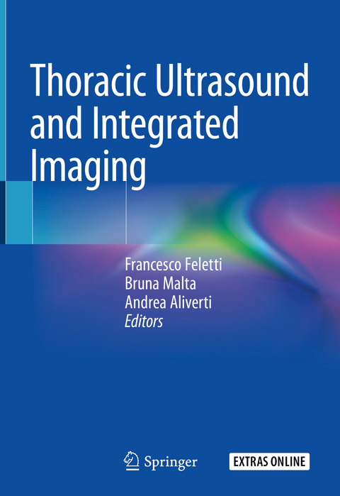
Thoracic Ultrasound and Integrated Imaging
Springer International Publishing (Verlag)
978-3-319-93054-1 (ISBN)
This book focuses on thoracic ultrasound, a versatile, diagnostically accurate, low-cost, noninvasive and non-ionizing imaging technique. Thanks to portable devices, the method can be used to provide quick and accurate diagnoses in emergency settings, during transport, or at the patient's bedside in intensive care units. In addition, as a dynamic examination that allows "real-time" assessment, it can be used to optimize diagnoses, the use of respiratory support equipment, surgical interventions and physiopathological assessments, both in critical patients and those with chronic conditions. Lastly, since it avoids ionizing radiation, thoracic ultrasound offers a first-line diagnostic tool for thoracic disease assessment in connection with pregnancy, neonatology and pediatrics.
Pursuing a practical approach, this book also addresses the technological components that are needed in order to adequately set up the equipment. This integrated approach provides non-radiologists with essential know-how on using thoracic ultrasound as an extension of their physical examinations. Specific chapters are dedicated to thoracic ultrasound applications in neonatology, pediatrics and emergency medicine, as well as guided procedures and diaphragm function studies. Thoracic ultrasound has been a central element in the editors' clinical and experimental work for several years, and the book also includes contributions by prominent international experts on specific applications. Given its content and scope, the book will be of interest to all medical practitioners seeking a practical approach to thoracic ultrasound.
Francesco Feletti is a Radiologist at the Local Health Authority of Romagna's Department of Diagnostic Imaging, at the S. Maria delle Croci Hospital in Ravenna. His main fields of practice are Emergency Radiology, Musculoskeletal Imaging and Interventional Imaging. In addition, he has a unique research record and considerable expertise in extreme sports medicine. Bruna Malta is a Radiologist at the University Hospital of Ferrara's Department of Diagnostic Imaging and Laboratory Medicine. Her main fields of interest are Diagnostic Imaging of the Thorax, Emergency Radiology and Pediatric Radiology. Andrea Aliverti is Full Professor, Dipartimento di Elettronica, Informazione e Bioingegneria (DEIB), Politecnico di Milano University, Italy. His main research fields include Bioengineering of the Respiratory System, Physiological Measurements, Biomedical Instrumentation, Lung Imaging, Functional Evaluation and Respiratory Mechanics.
Part I Technique.- Technological elements.- Artefacts in thoracic ultrasound.- Technical execution.- Part II Semeiotics and Integrated Imaging.- Lung consolidations.- Interstitial lung diseases.- Pleural conditions.- Chest wall disorders.- Mediastinal pathologies.- Part III Applications.- Neonatology.- Pediatrics.- Emergency medicine and intensive care.- Trauma.- Ultrasound guided procedures.- Diaphragm morphological and functional study.- Contrast medium in thoracic ultrasound.
"It is well written and illustrated and highly recommended as a text for aspiring and experienced practitioners, including sonographers, clinicians and radiologists." (D Maudgil, RAD Magazine, October, 2020)
| Erscheinungsdatum | 12.03.2020 |
|---|---|
| Zusatzinfo | VIII, 262 p. 456 illus., 313 illus. in color. With online files/update. |
| Verlagsort | Cham |
| Sprache | englisch |
| Maße | 178 x 254 mm |
| Gewicht | 937 g |
| Themenwelt | Medizinische Fachgebiete ► Radiologie / Bildgebende Verfahren ► Radiologie |
| Schlagworte | B lines • contrast agents • Pneumonia • Pneumothorax • Thoracic Surgery • ultrasound-guided procedures |
| ISBN-10 | 3-319-93054-0 / 3319930540 |
| ISBN-13 | 978-3-319-93054-1 / 9783319930541 |
| Zustand | Neuware |
| Informationen gemäß Produktsicherheitsverordnung (GPSR) | |
| Haben Sie eine Frage zum Produkt? |
aus dem Bereich


