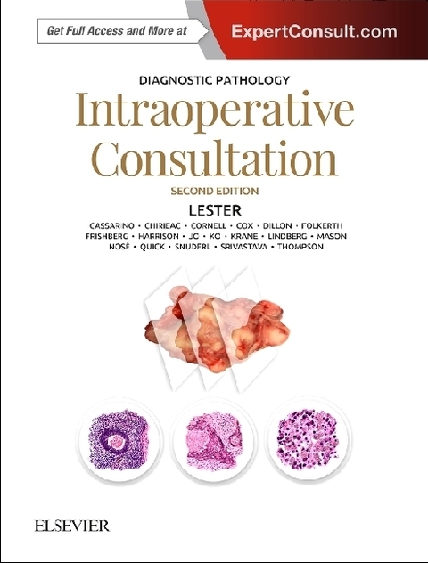
Diagnostic Pathology: Intraoperative Consultation
Elsevier - Health Sciences Division (Verlag)
978-0-323-57019-0 (ISBN)
- Separate chapters cover more than 60 questions posed by surgeons during intraoperative consultations, in addition to general introductory and methods chapters
- More than 1,700 outstanding images
- Time-saving reference features include bulleted text, tables of diagnostic criteria, annotated images, and an extensive index
- Thoroughly updated content in every chapter includes new criteria for diagnosis (e.g., limiting surgery for some types of lung lesions), more information on gross evaluation of specimens, and additional photographs for many diagnoses
- New chapters include new techniques, such as image-guided lung resections, finding and safely handling radioactive seeds for breast biopsies, tissue banking techniques, nipple margin evaluation for nipple-sparing mastectomy, and evaluation of specimens from patients with epilepsy
- Expert ConsultT eBook version included with purchase. This enhanced eBook experience allows you to search all of the text, figures, and references from the book on a variety of devices
Susan C. Lester, MD, PhD, is the Former Chief of Breast Pathology Services at Brigham and Women's Hospital and Associate Professor with Harvard Medical School in Boston, Massachusetts
General
Intraoperative Consultation: Introduction
Quality Assurance
Safety Precautions
Telepathology
Methods
Gross Examination
Cytologic Examination
Frozen Section
Slide Preparation
Tissue Allocation for Special Studies and Banking
Breast: Radioactive Seed Localization
Lymph Nodes: Molecular Methods for Evaluation
Contents
Adrenal and Paraganglia: Diagnosis
Anterior Mediastinal Mass: Diagnosis
Appendix: Diagnosis
Bone Lesion/Tumor: Diagnosis and Margins
Breast: Diagnosis
Breast: Parenchymal Margins
Breast: Nipple Margin Evaluation
Bronchus and Trachea: Diagnosis
Cerebellum and Brainstem: Diagnosis
Cerebral Hemispheres: Diagnosis
Cerebral Hemispheres: Evaluation for Epilepsy
Colon: Diagnosis and Margins
Colon: Evaluation for Hirschsprung Disease
Esophagus: Diagnosis and Margins
Fallopian Tube: Diagnosis
Head and Neck Mucosa: Diagnosis and Margins
Kidney, Adult: Diagnosis and Margins
Kidney: Evaluation of Allograft Prior to Transplantation
Kidney Needle Biopsy: Evaluation for Adequacy
Kidney, Pediatric: Indications and Utility
Larynx: Diagnosis and Margins
Liver, Capsular Mass: Diagnosis
Liver: Evaluation of Allograft Prior to Transplantation
Liver, Intrahepatic Mass: Diagnosis and Margins
Lung, Ground Glass-Opacities and Small Masses: Image-Guided Resection
Lung: Margins
Lung: Nonneoplastic Diffuse Disease: Diagnosis
Lung Mass: Diagnosis
Lymph Nodes, Axillary: Diagnosis
Lymph Nodes Below Diaphragm: Diagnosis
Lymph Nodes: Diagnosis of Suspected Lymphoproliferative Disease
Lymph Nodes, Head and Neck: Diagnosis
Lymph Nodes, Mediastinal: Diagnosis
Meninges: Diagnosis
Nasal/Sinus: Diagnosis of Suspected Fungal Rhinosinusitis
Nasal/Sinus: Diagnosis of Suspected Neoplasia
Oropharynx and Nasopharynx: Diagnosis
Ovary, Mass: Diagnosis
Pancreas, Biopsy: Diagnosis
Pancreas Resection: Parenchymal, Retroperitoneal, and Bile Duct Margins
Parathyroid Gland: Diagnosis and Margins
Peripheral Nerve and Skeletal Muscle: Allocation of Tissue for Special Studies
Peritoneal/Omental Mass: Biopsy
Pituitary: Diagnosis
Pleura: Diagnosis
Revision Arthroplasty: Evaluation of Infection
Salivary Gland: Diagnosis and Margins
Skin: Diagnosis and Margins
Skin: Evaluation for Toxic Epidermal Necrolysis vs. Staphylococcal Scalded Skin Syndrome
Skin: Mohs Micrographic Surgery
Soft Tissue: Evaluation for Necrotizing Fasciitis
Soft Tissue Mass: Diagnosis and Margins
Spinal Cord: Diagnosis
Stomach: Diagnosis and Margins
Thyroid: Diagnosis
Ureter: Margins
Uterus, Endometrium: Diagnosis
Uterus, Endometrium: Diagnosis of Pregnancy
Uterus, Myometrium: Diagnosis
Vulva: Diagnosis and Margins
| Erscheinungsdatum | 10.04.2018 |
|---|---|
| Reihe/Serie | Diagnostic Pathology |
| Zusatzinfo | Approx. 1400 illustrations (1400 in full color); Illustrations, unspecified |
| Verlagsort | Philadelphia |
| Sprache | englisch |
| Maße | 222 x 281 mm |
| Gewicht | 1700 g |
| Einbandart | gebunden |
| Themenwelt | Studium ► 2. Studienabschnitt (Klinik) ► Pathologie |
| ISBN-10 | 0-323-57019-4 / 0323570194 |
| ISBN-13 | 978-0-323-57019-0 / 9780323570190 |
| Zustand | Neuware |
| Informationen gemäß Produktsicherheitsverordnung (GPSR) | |
| Haben Sie eine Frage zum Produkt? |
aus dem Bereich


